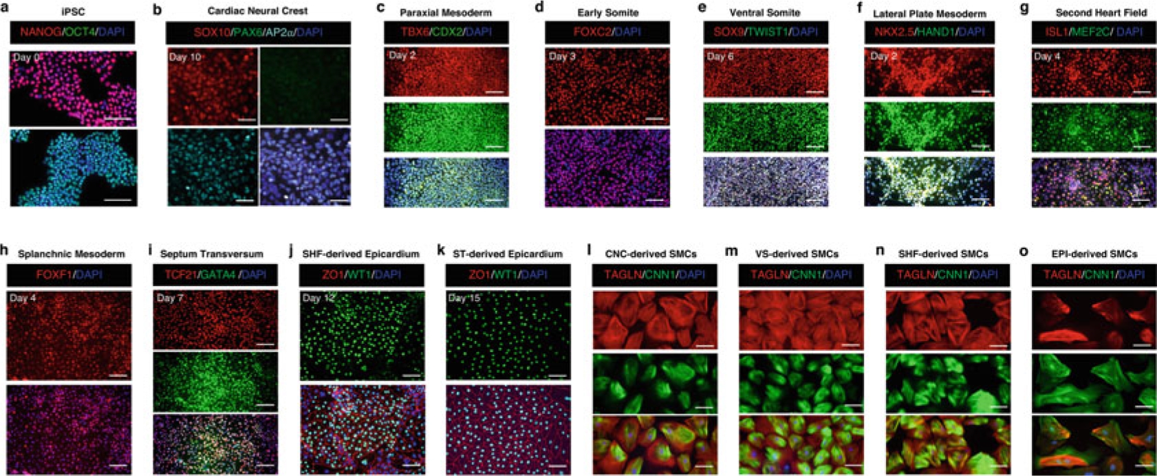
Fig. 2.

Representative immunofluorescence images showing phenotypic markers for specific cell types during embryonic origin-specific VSMC differentiation. (a–k) The time point labeled on each set of cell marker-positive fluorescence images is consistent with that shown on the bright-field image of the same cell type in Fig. 1. (l–o) Embryonic origin-specific VSMCs are positive for TAGLN and CNN1 after being cultured in the SMC growth medium for 14 days. Scale bars represent 100 μm
