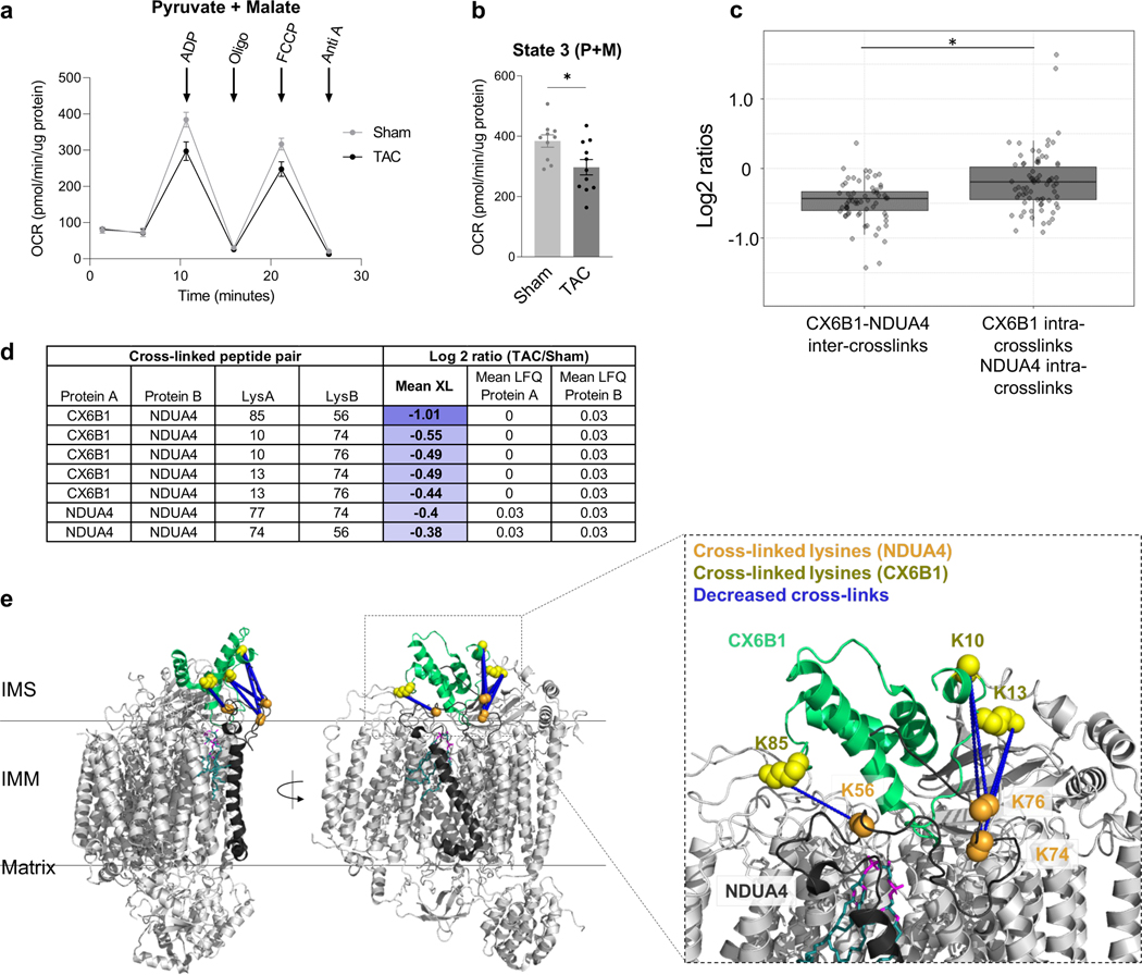
Figure 2: Decreased interaction between NDUA4 and C6XB1 affects CIV activity in TAC.
(a) Oxygen consumption rate of mitochondria isolated from TAC (n=11 animals) and Sham (n=10 animals) hearts given pyruvate/malate substrates followed by sequential injections of ADP, Oligomycin, FCCP, and Antimycin A, measured by Seahorse XFe24 Analyzer.
(b) State III-driven respiration of isolated mitochondria (after addition of ADP).
(c) Box plot illustrates a comparison of Log2 ratios of inter-links versus intra-links of NDUA4 and CX6B1 (n=66 inter-protein crosslinks, n=84 intra-protein crosslinks, visualized as median and 25th and 75th percentiles, with whiskers indicating minima and maxima excluding outliers). Welch’s two-sample, two-sided t-test was used to determine statistical significance.
(d) Table summarizing the mean cross-linking ratio and LFQ ratio for cross-linked peptide pairs exhibiting quantitative changes in NDUA4 and CX6B1 subunits of CIV.
(e) Monomeric Complex IV (grey, PDB: 5Z62). Interactions between NDUA4 (black) and CX6B1 (green) subunits are shown. Cross-linked lysine sidechains are shown as yellow (CX6B1) or orange (NDUA4) spheres. Cross-links exhibit a structural scaffold on the IMS-facing interface of CIV, which are decreased in TAC. Cardiolipin bound to CIV is shown in teal.
For (a-b), TAC (n=11 animals) and Sham (n=10 animals) data is represented as AVG+/−SEM, *denotes p<0.05 by unpaired, two-tailed Student’s t test.

