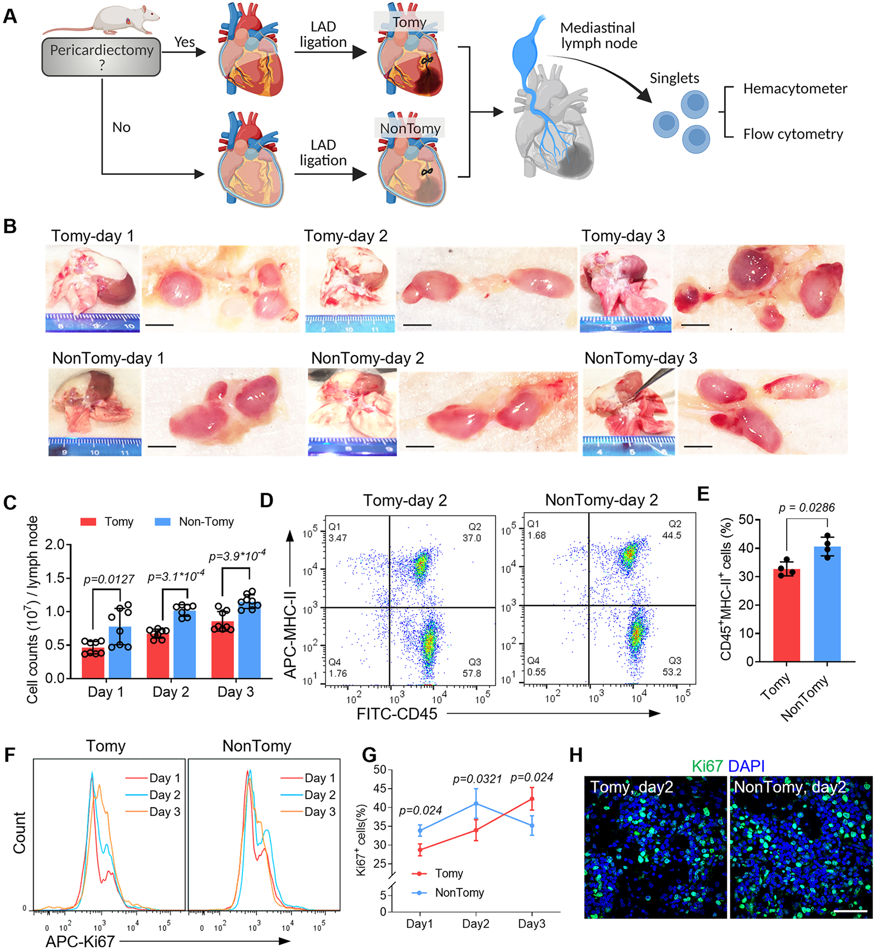
Figure 1. Pericardial drainage promotes immune activation in cardiac-draining mediastinal lymph node (MLN).

(A) Schematic study design. (B) Macroscopic examination of the cardiac-draining MLN. Scale bar, 1mm. (C) Cell counts in each MLN was measured by using hemacytometer. n=8 lymph nodes for Tomy group (Day 1-3) and NonTomy group (Day 1 and Day 3); n=7 lymph nodes for NonTomy group (Day 2). (D) Flow cytometry detection of APC migration in the MLN by using MHC-II as a marker. (E) Quantitative data of CD45+MHC-II+ cell counts in the MLN. n=4 animals for each group. (F) Flow cytometry detection of cell proliferation in the MLN, and accordingly the percentage of Ki67+ cells were plotted (G). n=4 animals for each group. (H) Immunofluorescent staining of Ki67 to show the cell proliferation in the MLN. Scale bar, 60μm. Quantitative data was shown as mean ± SD. p values were determined by one-way ANOVA for C; unpaired 2-tailed nonparametric Mann-Whitney test for E, G.
