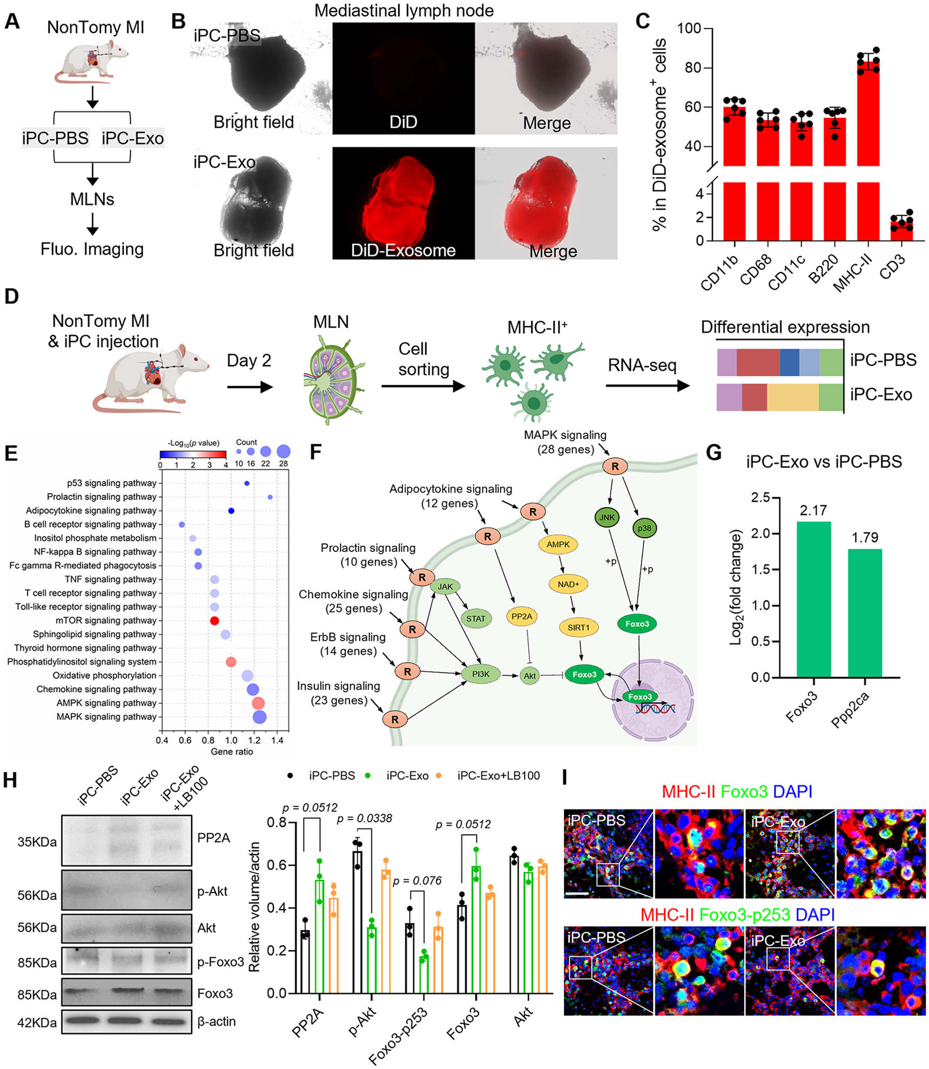
Figure 2. iPC injection of MSC-exosomes promotes Foxo3 activation in APCs.

(A) Schematic illustration of intrapericardial injection of MSC-exosomes for MI treatment. (B) Macroscopic fluorescent imaging to show the accumulation of MSC-exosomes in the MLN. (C) Flowcytometric detection of exosomes uptake by cells in the MLN. n=6 animals for each test. (D) Study design for RNA-seq analysis. (E) KEGG pathway enrichment analysis. (F) Integrated analysis of the enriched signal pathways. (G) Analysis of Foxo3 and PP2A(Ppp2ca) expression in RNA-seq data. The values were shown as relative expression compared to iPC-PBS group. (H) Western-blot detection of PP2A, p-Akt, Foxo3 and Foxo3-p253 in APCs. Quantitative data was acquired from three independent tests. (I) Immunostaining detection of Foxo3 and Foxo3-p253 expression in the MLN. Scale bar, 60μm. Quantitative data was shown as mean ± SD. p value was determined by Kruskal-Wallis’s test with Dunn’s correction for H.
