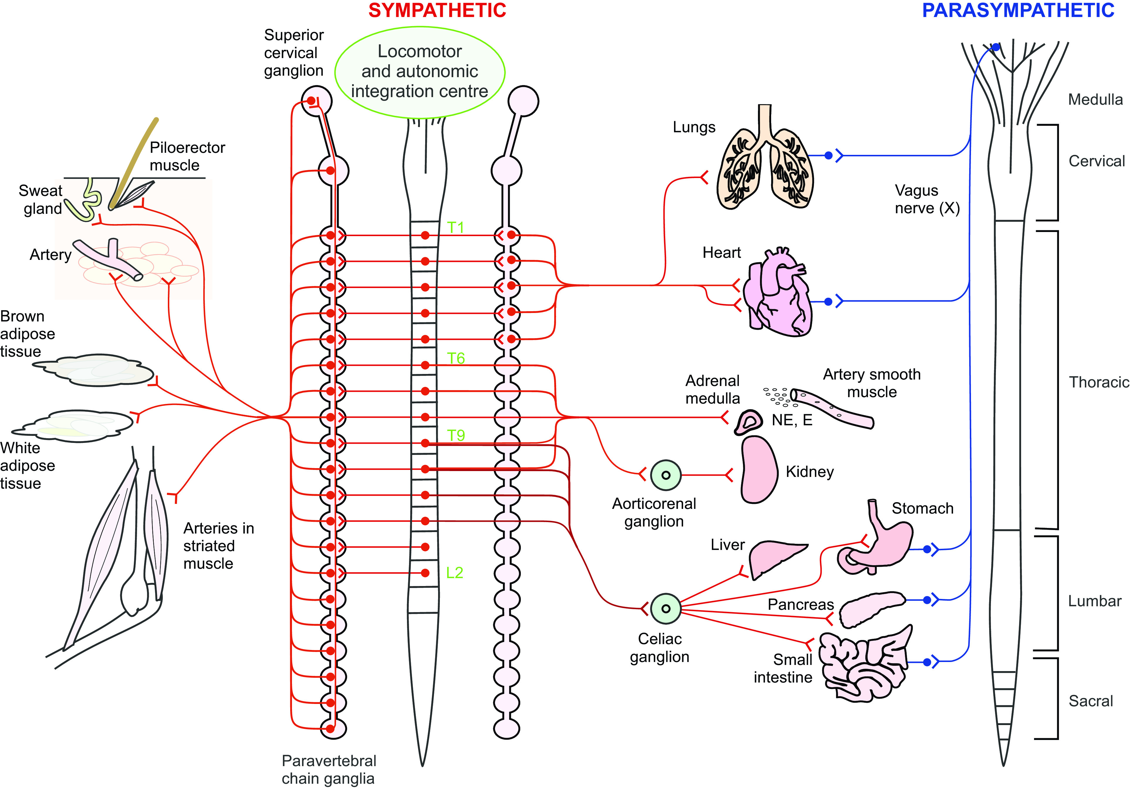
Figure 1.

Schematic of central and peripheral localization of preganglionic and postganglionic parasympathetic and sympathetic homeostatic and metabolic autonomic input to key tissues and organs supporting movement and exercise. Spinal levels providing sympathetic input to the lungs, heart, adrenal medulla, sweat glands, white and brown adipose tissue, and smooth muscle of arteries of the splanchnic regions and skeletal muscle tissue are shown in red. Organs that also receive parasympathetic input shown in blue. During movement and exercise parasympathetic input is reduced, limiting digestion, urine production etc., while sympathetic drive to these sympathetic targets is increased. Reprinted with permission from Cowley (8).
