Figure 1.

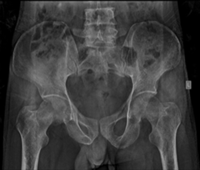
Anteroposterior radiograph of pelvis with bilateral hip demonstrating diffuse osteoporosis of the visualised pelvic bones and femur, with loss of continuity of the primary and secondary trabecula in the proximal femur bilaterally

Anteroposterior radiograph of pelvis with bilateral hip demonstrating diffuse osteoporosis of the visualised pelvic bones and femur, with loss of continuity of the primary and secondary trabecula in the proximal femur bilaterally