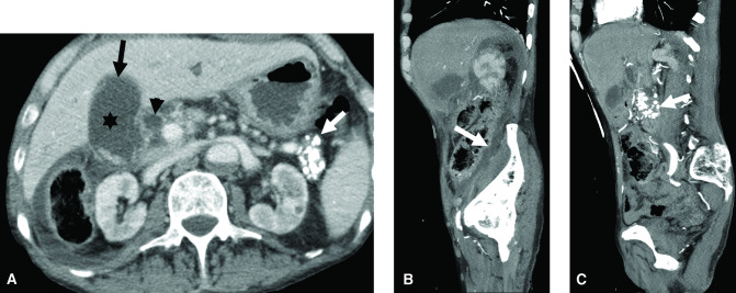
Figure 1.
Abdominal CT pre cholecystectomy showing gangrenous cholecystitis. Axial (A) and sagittal (B, C) images demonstrating changes of acute gangrenous cholecystitis. There is a distended gallbladder with layering stones (asterisk, A), mural discontinuity in the fundal region (black arrow, A), and small pericholecystic fluid. There is a small amount of free fluid (arrow, B) extending from the right upper quadrant to the right lower quadrant without evidence of retroperitoneal abscess. Also note the intra- and extrahepatic (arrowhead, A) biliary dilation, and extensive pancreatic calcifications consistent with chronic pancreatitis (white arrow, A, C).

