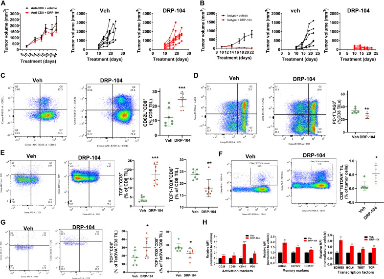
Fig. 8. DRP-104 causes CD8+ T cell–dependent tumor regression secondary to memory stem-like conditioning of infiltrating CD8+ T cells.
(A) MC38-bearing CES1−/− mice treated with DRP-104 (0.3 mg/kg DON equivalent, subcutaneously, 5 days per week) after depletion of CD8+ T cells; spider plots of individual mice data from vehicle or DRP-104. (B) OVA-expressing MC38-bearing CES1−/− mice treated with vehicle or DRP-104; tumor growth curves of individual mice are depicted from vehicle and DRP-104 groups. Representative flow cytometry plots (left) and data charts (right) from CD8+ TIL showing (C) CD44 and CD62L, (D) PD-1 and LAG3, and (E) TCF1 and TOX defined subsets. (F) Flow cytometry showing antigen-specific tetramer-positive CD8+ TIL and (G) TCF1 and TOX defined subsets within tetramer-OVA–positive population. (H) Relative mean fluorescence intensity data plots for activation markers, memory/stem cell markers, and transcription factors in CD8 TILs. *P < 0.05, **P < 0.01, ***P < 0.001, and ****P < 0.0001, based on t test.

