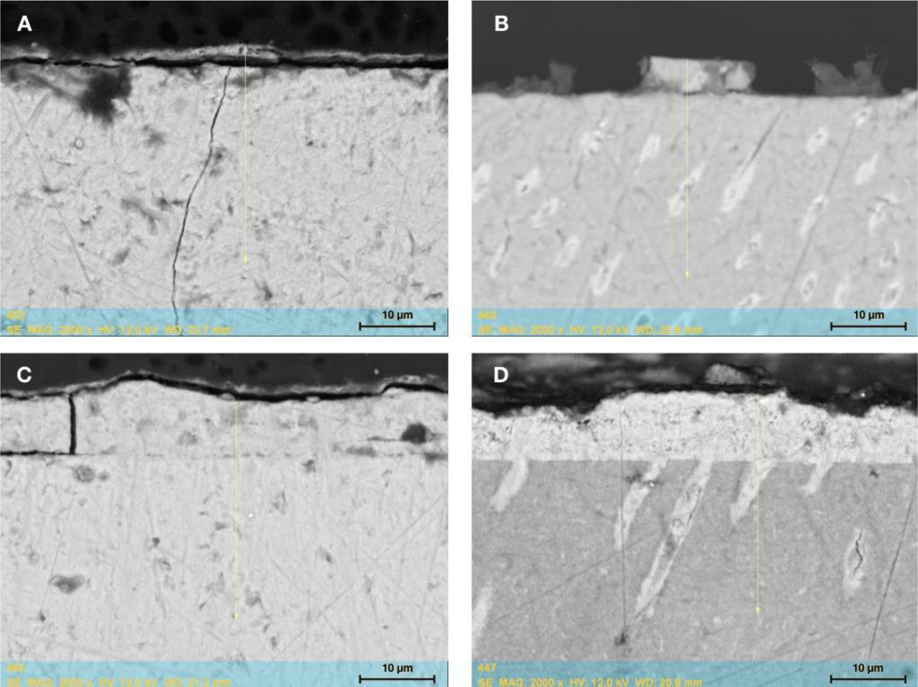
Figure 1:

SEM of representative enamel (A, C) and dentin samples (B, D). The enamel-like layer is visible on the surface of the treated samples. In dentin, the new mineral is also detectable in the tubules. Figures show the results after one (A, B) and five repeated applications (C, D) of BIMIN.
