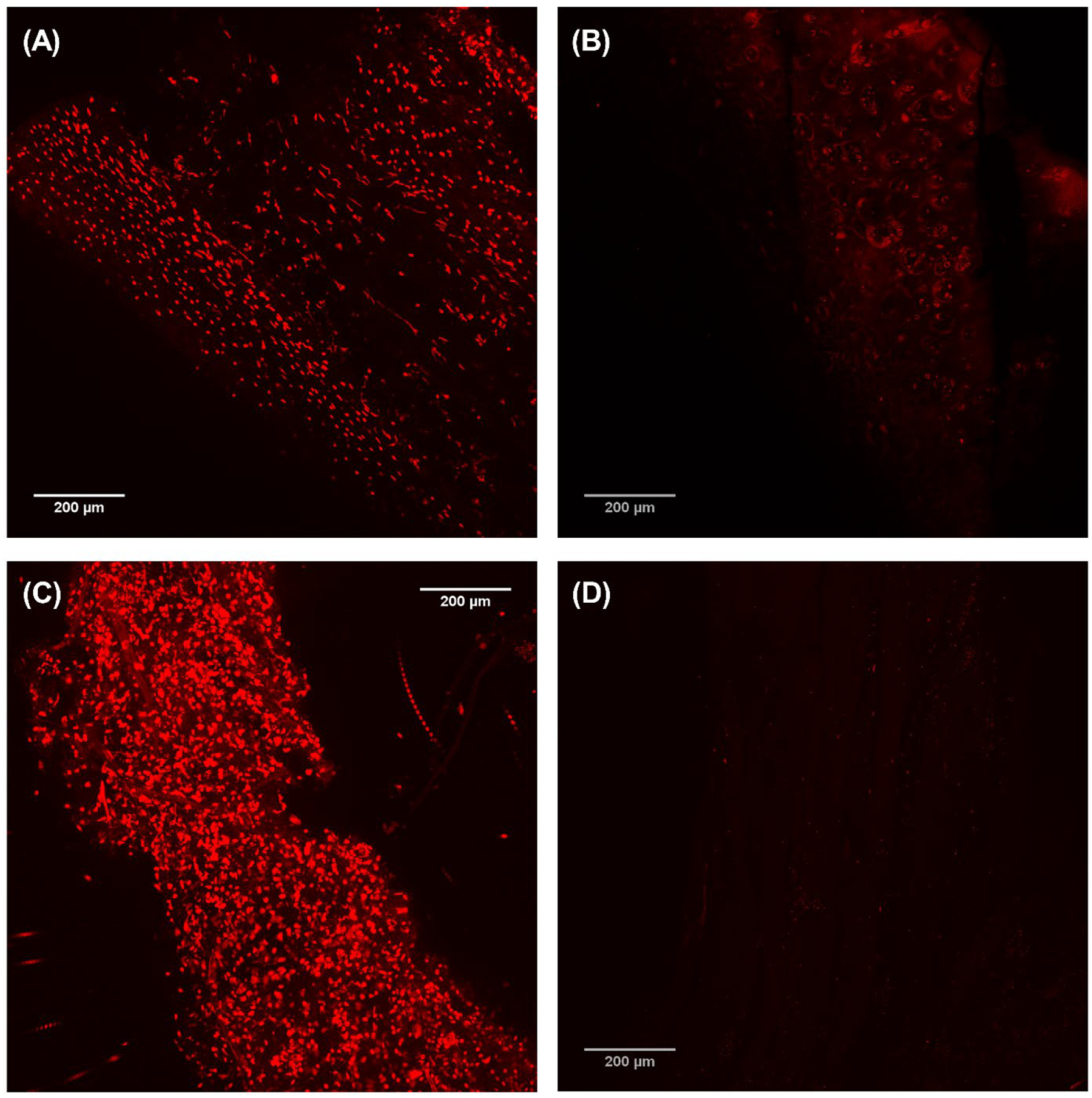
Figure 5:

Confocal microscopy of LIVE/DEAD™ stained samples of Larynx A. Images (A) and (B) show the cartilage before and after decellularization, respectively. Images (C) and (D) show the muscle before and after decellularization, respectively. Scale bar = 200 μm.
