
Fig. 1. Sperm chromatin is aggregated and contains a specific histone variant: H2B.8.
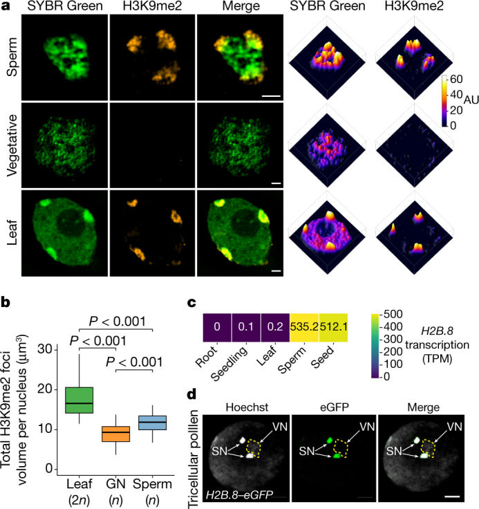
a, Super-resolution 3D-SIM images (left) and associated intensity profiles (right) of wild-type (WT) sperm and vegetative nuclei from pollen and a diploid leaf nucleus. DNA was stained with SYBR Green (green) and H3K9me2 was immunolocalized (orange). Data shown represent three independent experiments. AU, arbitrary units. b, Total volume of H3K9me2-enriched heterochromatin foci in a diploid leaf nucleus, generative nucleus (GN) and sperm nucleus. P values calculated using one-sided analysis of variance (ANOVA) followed by individual two-sample Tukey tests; n = 30 nuclei each examined over two independent experiments. All the boxplots in this work show median (thick black bar) and first and third quartiles, with lower and upper whiskers extending to 1.5-times the interquartile range of the first and third quartiles or the highest and lowest values, respectively. c, H2B.8 transcription levels in indicated tissues and cells. TPM, transcripts per million. d, Confocal images of pH2B.8::H2B.8–eGFP pollen, in which the eGFP signal is specific to the sperm nuclei (SN). VN, vegetative nucleus (outlined in a dashed line). Data shown represent three independent experiments. Scale bars, 1 μm (a) 5 μm (d).