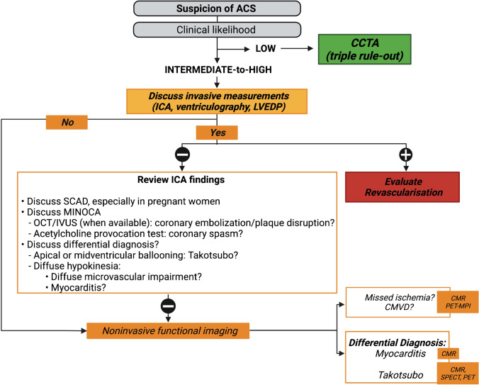
Fig. 1.
Proposed diagnostic algorithm for acute coronary syndrome. In case a STEMI is suspected, urgent ICA is recommended [235]. In case a NSTEMI is suspected, the diagnostic approach depends on the clinical likelihood of CAD, ECG findings, and troponin measurement [25]. If the clinical likelihood is low, ECG-triggered contrast-enhanced CT can rule out simultaneously coronary stenosis, aortic dissection, and pulmonary embolism (triple rule-out) [25]. If the clinical likelihood is intermediate-to-high, ICA must be discussed, urgently or semi-urgently. If no coronary stenosis is found on ICA, MINOCA should be suspected. A first step consists of thoroughly reviewing the ICA to search for subtle signs of SCAD, coronary embolization, or plaque disruption, using IVUS or OCT, when available [32, 236]. After symptom resolution and exclusion of other causes, invasive provocative testing using acetylcholine, ergonovine, or methylergonovine can help to establish a definitive diagnosis. Nevertheless, it should be used with caution and only by experienced operators, and in all cases never in the acute setting of the episode [29]. LV angiography can also provide important information such as segmental hypokinesia suggesting epicardial abnormality, apical or midventricular ballooning being in favor of TTC cardiomyopathy, and a more diffuse hypokinesia sometimes suggesting a microvascular impairment [237]. If a diagnosis cannot be established, advanced noninvasive imaging is required. CMR can show patterns of ischemia/infarct, evidence MINOCA causes [82, 83], and rule out differential diagnosis such as TTC and myocarditis [26, 30, 83]. MPI can also be used in the acute/subacute setting, after symptom resolution and normalization of ECG and troponin [238]. Abbreviations: ACS: acute coronary syndrome; CAD: coronary artery diseases; CMR: cardiac magnetic resonance; CCTA: coronary computed tomography angiography; CT: computed tomography; ECG: electrocardiogram; ICA: invasive coronary angiography; IVUS: intravascular ultrasound ; LV: left ventricle; LVEDP: left ventricular end-diastolic pressure; MI: myocardial infarction; MINOCA: myocardial infarction with no obstructive coronary artery disease; MPI: myocardial perfusion imaging; N: normal; NSTEMI: non ST-elevation myocardial infarction; OCT: optical coherence tomography; PET: positron emission tomography; SCAD: spontaneous coronary artery dissection; SPECT: single-photon emission computed tomography; STEMI: ST-elevation myocardial infarction; TTC: Takotsubo cardiomyopathy

