Case 4.
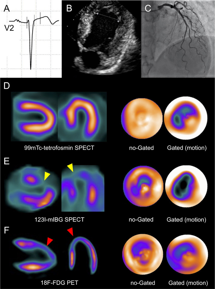
Sixty-year-old woman for routine checkup. A 60-year-old woman with severe hypercholesterolemia presented to the cardiology outpatient clinic for a routine evaluation. The resting ECG (A, V3 lead) showed a loss of R-wave progression and biphasic T-waves in the anterior precordial and lateral leads, respectively. Echocardiography (B) revealed isolated apical akinesia with preserved LVEF. Subsequent ICA was normal (C). ECG-gated 99mTc-MPI-SPECT performed 7 days later showed normal LV stress/rest perfusion, but reduced end-systolic thickening and antero-apical hypokinesia (D, horizontal and vertical long axes, polar maps). TTC was hypothesized and, 2 weeks after the perfusion scan, the patient underwent ECG-gated 123I-MIBG SPECT (E) and 18F-FDG PET/CT (F), showing a concordant alteration of adrenergic innervation (E, yellow arrowheads) and glucose metabolism (F, red arrowheads) in apical and anterior LV wall. A careful patient questioning revealed significant stress exposure at work triggering the (clinically silent) TTC. The resolution of the workplace conflict coincided with the recovery of LV apical kinetics documented by echocardiography 3 months later. TTC is often caused by severe emotional stress. If segmental LV wall motion disorders are present, an adrenergic cause must be considered. In this context, nuclear medicine provides a specific imaging pattern, useful to reach the diagnosis. Abbreviations: 18F-FDG: Fluor-18- radiolabeled fluorodeoxyglucose; 99mTc: 99mTechnetium; 123I-MIBG: 123I-meta-iodobenzylguanidine; CT: computed tomography; ECG: electrocardiogram; ICA: invasive coronary angiography; LV: left ventricle; LVEF: left ventricular ejection fraction; MPI: myocardial perfusion imaging; PET: positron emission tomography; SPECT: single-photon emission computed tomography; TTC: Takotsubo cardiomyopathy

