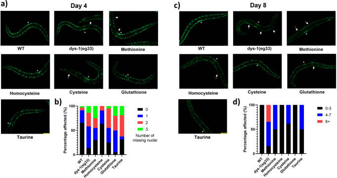
Fig. 3. SAA mildly delay onset of cell death at Day 4 post-adulthood, with significant preservation at Day 8.
a Representative images of muscle cell death in CB5600 (wt), CC91 (dys-1(eg33)) and CC91 treated with SAA: L-Methionine (10 mM), L-homocysteine (10 µM), L-cysteine (10 µM), L-glutathione (100 µM) and L-taurine (10 µM). Muscle cell death (as identified by the absence of muscle nuclei) was more pronounced in the dys-1(eg33) animals compared to wt. b The scatter bar graph shows a higher proportion of animals with no missing nuclei when treated with SAA bar L-glutathione. However, the proportion of animals with 2 or more missing nuclei was similar across all groups bar L-homocysteine. c Representative images of muscle cell death in CB5600 (wt), CC91 (dys-1(eg33)) and CC91 treated with SAA: L-Methionine (10 mM), L-homocysteine (10 µM), L-cysteine (10 µM), L-glutathione (100 µM) and L-taurine (10 µM). Muscle cell death was more pronounced in the dys-1(eg33) animals compared to wt at Day 8 post-adulthood. d The scatter bar graph shows a higher proportion of animals with 0–3 missing nuclei when treated SAA in particular, L-homocysteine and L-glutathione. Vulva is identified by the * and arrows show the missing nuclei. Scale bar: 30 µm. Data are means from 20 animals across two biological repeats.

