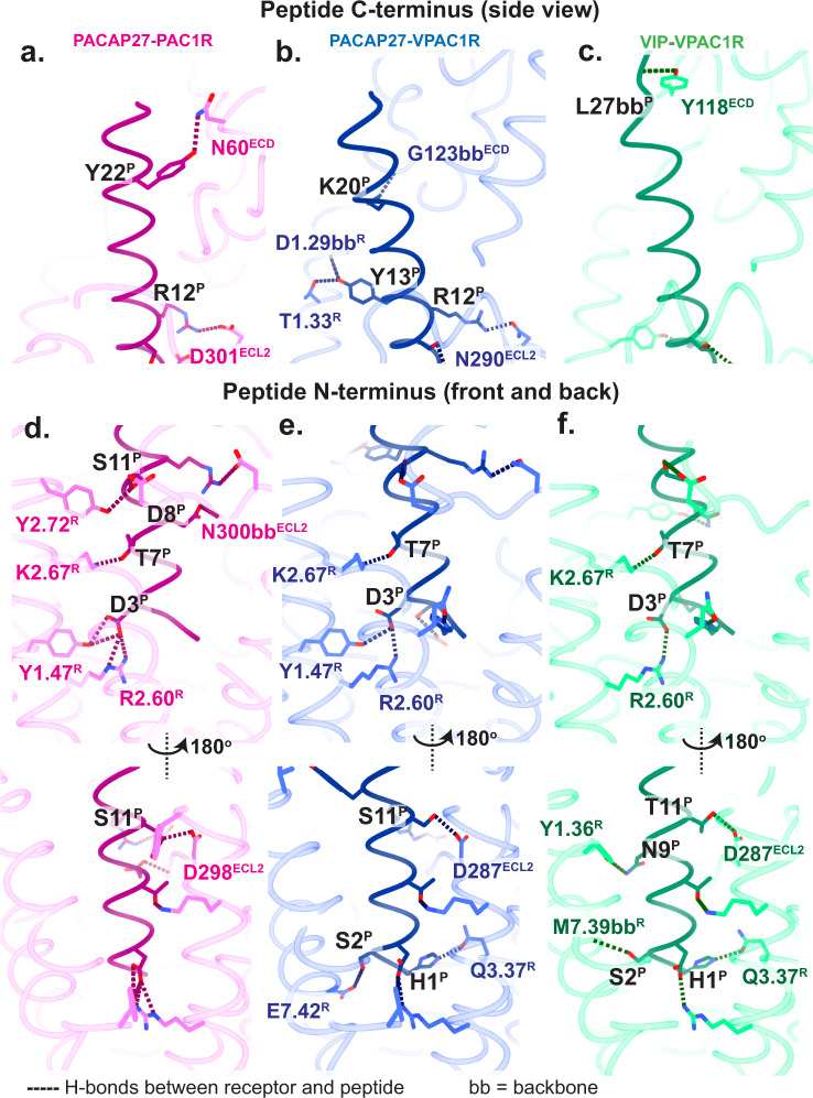
Fig. 2. Hydrogen bonds between peptide (P) and receptor (R) residues in the static structures.
The receptor backbone is displayed in transparent in ribbon format and peptide backbone in ribbon format, with sidechains that are involved in H-bonds displayed in stick format. PAC1R-PACAP27 is shown in pink-dark pink, VPAC1R-PACAP27 in blue-dark blue and VPAC1R-VIP in green-dark green. a–c Interactions of the peptide C-terminus and the receptor shown as side view. d–f Interactions of the peptide N-terminus and the receptor shown as front and back view. H-bonds between peptide and receptor are displayed as dotted lines. H-bonds involving backbones, and not sidechains, are labelled as ‘bb’. Receptor residues are numbered according to the Wootten et al.18, class B1 scheme. Residues in the ECL and ECD as well as peptide residues are labelled according to the residue number.

