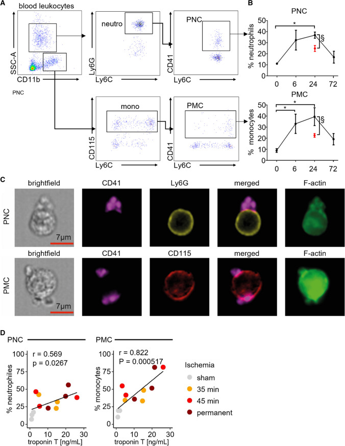
Fig. 1.
Platelet–leukocyte complexes surge in peripheral blood post-myocardial ischemia and reperfusion injury (I/R). A Representative dot plots depicting blood neutrophils, monocytes, platelet–neutrophil complexes (PNC), and platelet–monocyte complexes (PMC), respectively. B Time course for PNC (top) and PMC (bottom) fractions in blood before (t = 0) and after myocardial ischemia (35 min) with reperfusion over 72 h (black data points). *p < 0.05 denote statistically significant changes before vs after myocardial I/R (*) n = 4 per time point, Kruskal–Wallis test, Dunn’s multiple comparison, §p < 0.05 denote statistically significant changes between myocardial I/R (black) and sham (red) at 24 h of reperfusion, n = 4 per group, Mann–Whitney. C Representative ImageStreamX image PNC and PMC defining platelets as CD41 + , neutrophils as Ly6G + , monocytes as CD115 + and F-actin enrichment at the contact zone. D Correlation of plasma troponin and blood PMC or PNC levels following 35 min and 45 min of myocardial ischemia with reperfusion or permanent LAD ligation 24 h post-surgery. *p < 0.05 denote statistically significant changes, determination of correlation by Pearson’s r

