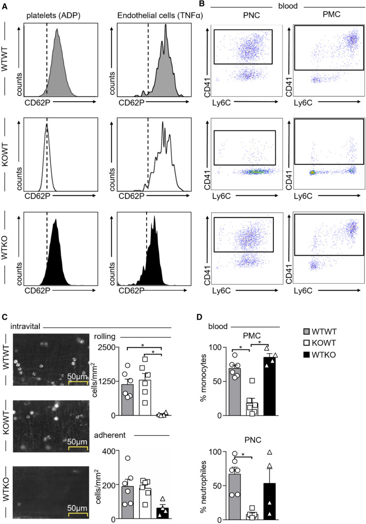
Fig. 2.
Surface expression of P-selectin (CD62P) and formation of platelet–leukocyte complexes in irradiated bone marrow chimeras, and its influence on leukocyte rolling and adhesion. Wild-type (WT) and P-selectin-deficient (KO) mice were lethally irradiated and reconstituted with either WT bone marrow in WT recipients (WTWT), KO bone marrow in WT recipients (KOWT), or WT bone marrow in KO recipients (WTKO). A Representative histograms showing selective loss of CD62P expression on platelets in KOWT chimeras and loss of CD62P expression on endothelial cells in WTKO chimeras. B Representative dot plots showing that selective loss of CD62P on platelets in KOWT chimeras reduces both platelet–neutrophil (PNC) and platelet–monocyte complexes (PMC) in blood. C Intravital microscopy of mesenteric venules in WTWT, KOWT, and WTKO chimeras 4 h post-intraperitoneal (i.p.) TNFα (200 ng) injection. Representative images of rhodamine-labeled leukocytes within the venules are shown on the left. Quantification of rolling and adherent leukocyte numbers are shown on the right. Results are presented as mean ± SEM, n = 6 for WTWT, KOWT and n = 4 for WTKO. *p < 0.05 denote statistically significant changes between groups as indicated using Kruskal–Wallis with Dunn’s multiple comparison test. D Quantification of blood PMC and PNC in WTWT, KOWT, and WTKO chimeras post-TNFα i.p. stimulation. Results are presented as mean ± SEM, n = 6 WTWT and KOWT, n = 4 WTKO. *p < 0.05 denote statistically significant changes between groups using Kruskal–Wallis with Dunn’s multiple comparison test

