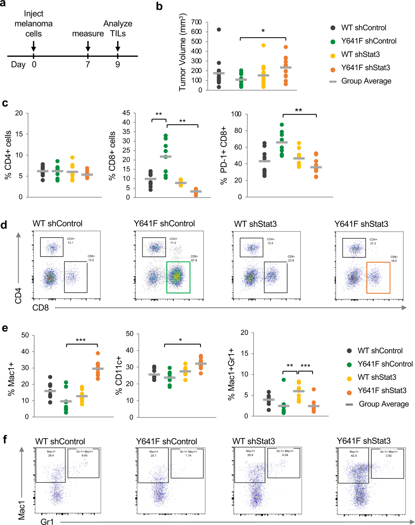
Figure 6. Assessment of tumor growth and tumor-infiltrating immune cells in Ezh2WT vs Ezh2Y641F melanoma cells.

(a) Schematic of experimental design. Melanoma cells were injected subcutaneously into the flanks of C57Bl/6 mice. Tumor dimensions were measured 7 days later, and tumors were collected on day 9 to analyze tumor-infiltrating lymphocytes (TILs). N = 8–10/group.
(b) In vivo tumor volume of Ezh2WT vs Ezh2Y641F melanoma cell lines, with and without Stat3 knockdown (*p<0.05).
(c) Analysis of tumor-infiltrating lymphocytes by flow cytometry, focusing on CD4+, CD8+ and CD8+/PD-1+ cells (**p<0.01).
(d) Representative flow cytometry plots for CD4+ and CD8+ cells from panel (c).
(e) Analysis of tumor-infiltrating myeloid cells by flow cytometry, using antibodies for Mac1, CD11c, and Gr1 (*p<0.05, **p<0.01, ***p<0.001).
(f) Representative flow cytometry plots for Mac1+ and Mac1+/Gr1+ cells from panel (e).
