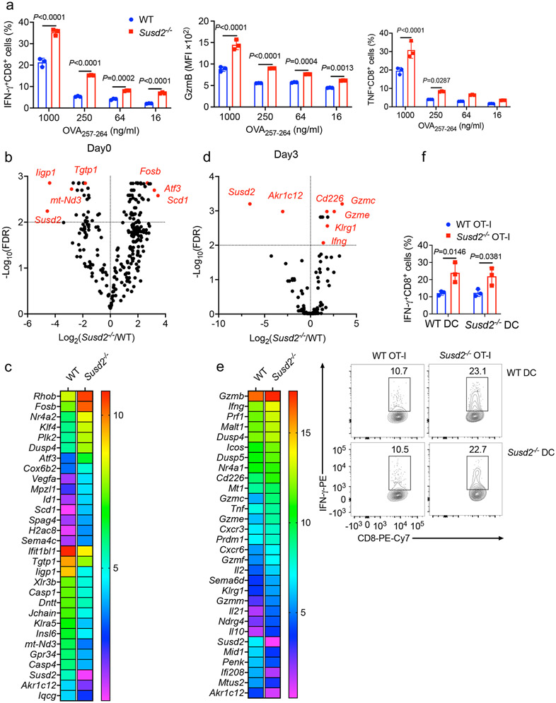
Extended Data Fig. 3. Susd2−/− CD8+ cells exhibit increased antitumor effector function.
a, Flow cytometry analysis of IFN-γ+CD8+ T cells, GzmB+CD8+ T cells and TNF+CD8+ T cells in different OVA257-264 dosage stimulated splenocytes isolated from WT or Susd2−/− OT-I mice. b-e, CD8+ T cells were isolated from total splenocytes of either WT or Susd2−/− OT-I mice left untreated or stimulated with OVA257-264 for 3 days and were subjected to RNA-seq assay. The volcano plot of RNA-seq data demonstrates differential gene expression between WT and Susd2−/− CD8+ T cells at Day 0 (b) and Day 3 (d). A heatmap of the top thirty genes representing genes differentially expressed between WT and Susd2−/− CD8+ T cells at Day 0 (c) and Day 3 (e). f, Intracellular accumulation of IFN-γ in CD8+ T cells isolated from WT or Susd2−/− OT-I mice that were cocultured with either WT or Susd2−/− bone marrow-derived dendritic cells (BMDCs) that have been pulsed with OVA257-264. a,f, n = 3. b-e, n = 4. n, number of mice per group. a,f, data are representative of four independent experiments. b-e, data are representative of two independent experiments. b,d statistical significance was calculated using two-sided Wilcoxon’s rank-sum test and adjusted with Bonferroni’s correction. Statistical significance was determined by two-way ANOVA followed by Sidak’s multiple comparisons test(a,f) with P values noted in the figure. All data are mean ± SD.

