
Fig. 1 |. The pancreas is innervated by neurons in coeliac, nodose, dorsal root and intrapancreatic ganglia.
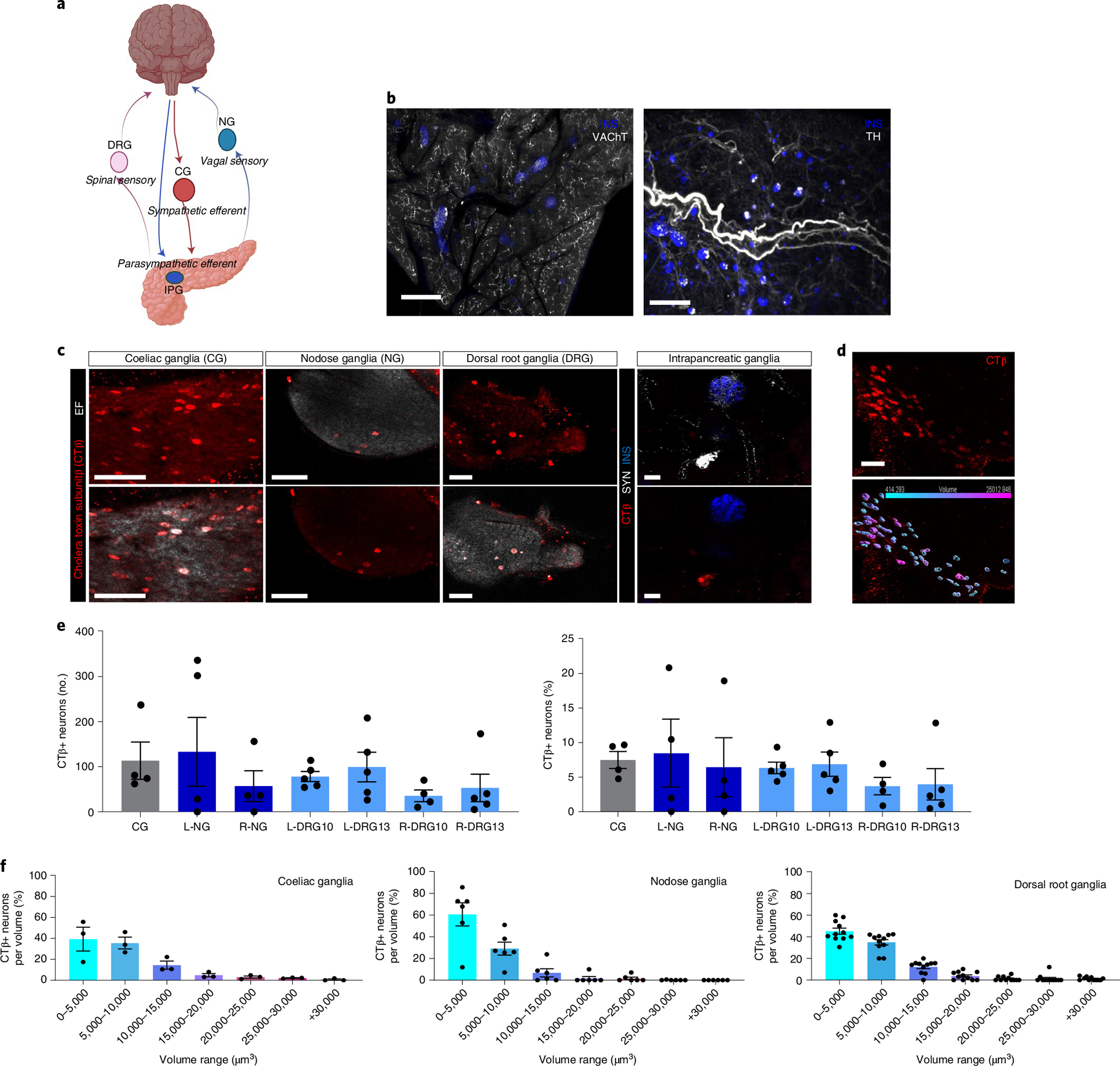
a, Schematic representation of pancreas innervation. b, Maximum projections of lightsheet microscopy images of mouse pancreatic samples cleared with iDISCO+ and stained for insulin (blue) and vesicular acetylcholine transporter, VAChT (white, left), or tyrosine hydroxylase, TH (white, right). Scale bars, 300 μm. c, Representative confocal images of CTβ+ pancreas-innervating neurons (red, top row) in cleared peripheral ganglia (CG, NG, DRG and IPG) after intrapancreatic injection. Bottom row shows CTβ+ pancreas-innervating neurons (red) and endogenous fluorescence (EF) (white) for CG, NG and DRG. Upper right panel shows synapsin (SYN) marking intrapancreatic ganglia (white) and insulin (blue), bottom right panel shows CTβ+ pancreas-innervating neurons (red) and insulin (blue). Scale bars, 100 μm. d, Representative segmentation of CTβ+ pancreas-innervating neurons in CG showing CTβ+ pancreas-innervating neurons (red, top left side) and 3D volumes colour-coded for size (bottom left side). Two independent studies (3 and 2 ganglia, respectively). Scale bar, 100 μm. e, Quantification of CTβ+ pancreas-innervating neurons as total number per ganglion (left) and percentage of the total neurons per ganglion (number of CTβ+ pancreas-innervating neurons in specified ganglia/total number of neurons in specified ganglia) (right) after intrapancreatic injection of CTβ. Data are shown for left and right DRG at T10 and T13 (L-DRG10, L-DRG13, R-DRG10 and R-DRG13). Biologically independent sample numbers: CG, 4 samples; L-NG, 4 samples; R-NG, 4 samples; L-DRG10, 5 samples; L-DRG13, 5 samples; R-DRG10, 4 samples; and R-DRG13, 5 samples. f, Volume distribution of CTβ+ pancreas-innervating neurons within each ganglion in CG (left), NG (middle) and DRG (right) (N = 3 mice). All data are represented as mean ± SEM. Individual data points represent individual ganglia. Statistical analyses are described in Supplementary Table 2. Figure 1a was created with BioRender.com.