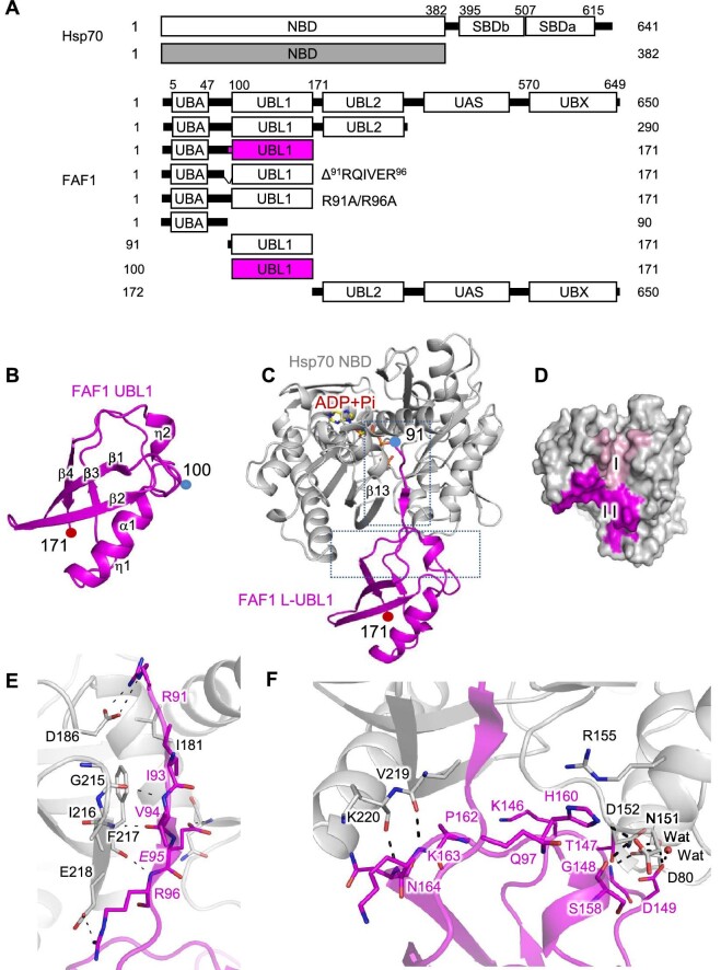
Figure 1.
Crystal structures of FAF1 UBL and FAF1 L-UBL1 complexed with Hsp70 NBD. (A) Domain structures of FAF1 and Hsp70 and the constructs prepared in this study. (B) Ribbon presentation of FAF1 UBL1 alone (aa 100–171). The secondary structures are indicated. (C) Ribbon representation of the complex of FAF1 L-UBL1 (aa 91–100) and Hsp70 NBD (aa 1–362) shown in magenta and silver, respectively, with the bound adenosine diphosphate (ADP) and phosphate (Pi) shown in the stick model. Subdomains of Hsp70 are indicated. (D) The molecular surface of Hsp70 within 4.0 Å from FAF1 is colored. Residues in contact with the linker region of FAF1 (aa 91–99) are colored in pink, while residues in contact with the UBL1 domain (aa 100–171) are in magenta. (E and F) Interactions between FAF1 L-UBL1 and Hsp70 NBD are shown in the stick model. Hydrogen bonds are indicated as dashed lines.

