Figure 3.
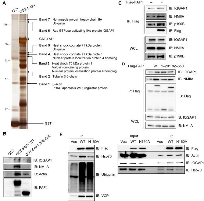
FAF1 forms a complex with IQGAP1 and actomyosin. (A and B) GST and GST-FAF1 (A) or GST, GST-FAF1 WT, and GST-FAF1 (aa 362–650) (B) were incubated with glutathione-agarose beads for 3 h and washed six times with washing buffer. GST-fused protein-bound beads were further incubated with HeLa cell lysates for 3 h, and then the beads were washed six times. Protein complexes were separated by SDS–PAGE and detected by silver staining (A) or analysed by western blotting (B). (C) HeLa cells were transfected with Flag or Flag-FAF1. After 24 h, cell lysates were immunoprecipitated with anti-Flag antibody, and the immune complex was analysed by western blotting. (D) HeLa cells were transfected with Flag, Flag-FAF1 WT, Flag-FAF1 (aa 1–201), or Flag-FAF1 (aa 82–650). After 24 h, cell lysates were immunoprecipitated with anti-Flag antibody, and the immune complex was analysed by western blotting. (E) HeLa cells were transfected with FAF1 WT or FAF1H160A mutant and then immunoprecipitated with anti-Flag antibodies. Immunoprecipitated proteins were immunoblotted with the indicated antibodies.

