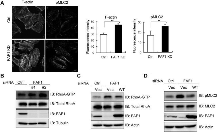
Figure 4.
FAF1 silencing promotes actin stress fiber formation and FAF1 negatively regulates RhoA activation. (A) HeLa cells transfected with control siRNA or FAF1 siRNA#2 for 72 h were fixed and stained for F-actin (phalloidin) or double phosphorylated MLC2 (Thr18/Ser19) to investigate stress fiber formation. The bar graph indicates the fluorescence intensities for F-actin or double phosphorylated MLC2 in the left panel. The experiments were conducted in quadruplet. Scale bar, 10 μm. (B) HeLa cells were transfected with two types of FAF1 siRNAs, #1 and #2. After 72 h, cells were lysed, and the lysates were incubated with GST-RBD of Rhotekin to measure the level of GTP-bound RhoA. Cell extracts were used to analyse the amount of total RhoA by western blotting. The relative amount of RhoA-GTP in control and FAF1 KD cells was determined by western blotting followed by densitometry analysis. (C) HeLa cells with silenced FAF1 (by transfecting with control siRNA or FAF1 siRNA#2 for 48 h) were transfected with Flag or Flag-FAF1 as indicated for adding back FAF1. At 24 h post-transfection, cells were lysed, and the lysates were incubated with GST-RBD of Rhotekin to measure the level of GTP-bound RhoA. Cell extracts were analysed using the indicated antibody. The relative amount of RhoA-GTP was determined by western blotting followed by densitometry analysis. (D) HeLa cells were transfected with control siRNA or FAF1 siRNA#2 for 48 h and transfected with Flag or Flag-FAF1 as indicated. At 24 h post-transfection, cells were lysed with gel sample buffer and analysed using the indicated antibodies.

