Figure 1.
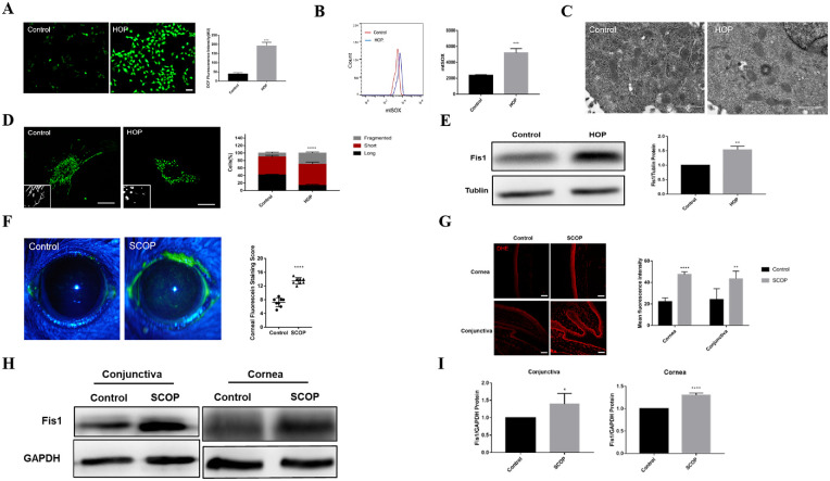
Hyperosmolarity induced mitochondrial oxidative damage and mitochondrial fission in vitro and in vivo. Some immortalized HCECs were treated with hyperosmotic medium and some served as controls. (A) H2DCFDA fluorescence in the control and HOP groups. Scale bar: 50 µm. (B) Flow cytometry revealed the mitochondrial ROS (mtROS) level in untreated (control) and HOP-stressed HCECs. (C) TEM image of mitochondrial structure in HCECs after exposure to HOP or normal medium. Scale bar: 1 µm. (D) Images showing mitochondrial morphology of HCECs under the confocal microscope and quantitative analysis of fragmented mitochondria. Scale bar: 20 µm. (E) Western blot results showing the changes in Fis1 expression in untreated and HOP-stressed primary HCECs. Experimental mice were injected subcutaneously with SCOP for 5 days and placed in a dry environment. (F) Representative corneal staining images and mean corneal staining scores of the normal and dry eye mice. (G) Dihydroethidium (DHE) fluorescent staining and quantitative analysis of the corneal and conjunctival epithelium of normal and dry eye mice. Scale bar: 50 µm. (H) Western blot results showing Fis1 expression in the corneal epithelium of normal and dry eye mice. (I) Quantitative analysis of Fis1 protein expression. Each group had five mice. *P < 0.05, **P < 0.01, ***P < 0.001, ****P < 0.0001.

