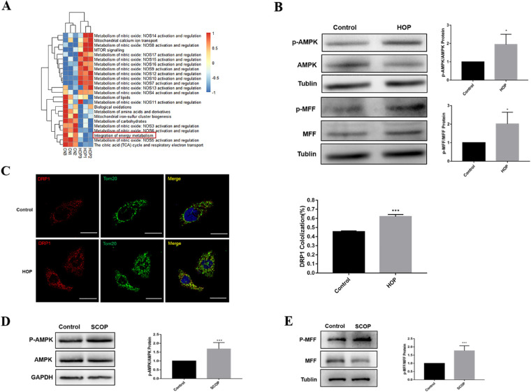
Figure 3.
The AMPK/MFF/DRP1 pathway was activated in vitro and in vivo. (A) The differentially expressed gene sets between the control and HOP groups. (B) Western blot results showing the changes in AMPK, p-AMPK, MFF, and p-MFF expression after 36 hours in HCECs exposed to hyperosmotic medium relative to that in normal medium. (C) Immunofluorescence analysis of DRP1 and Tom20 co-localization in HCECs after HOP for 36 hours. Scale bar: 20 µm. Cellular results are presented as the mean ± SD of three independent experiments. (D) Western blot results showing AMPK and p-AMPK expression of the conjunctiva in normal and dry eye mice. (E) Western blot was conducted to detect MFF and p-MFF expression of the conjunctiva in normal and dry eye mice, five mice in each group. *P < 0.05,**P < 0.01, ***P < 0.001.

