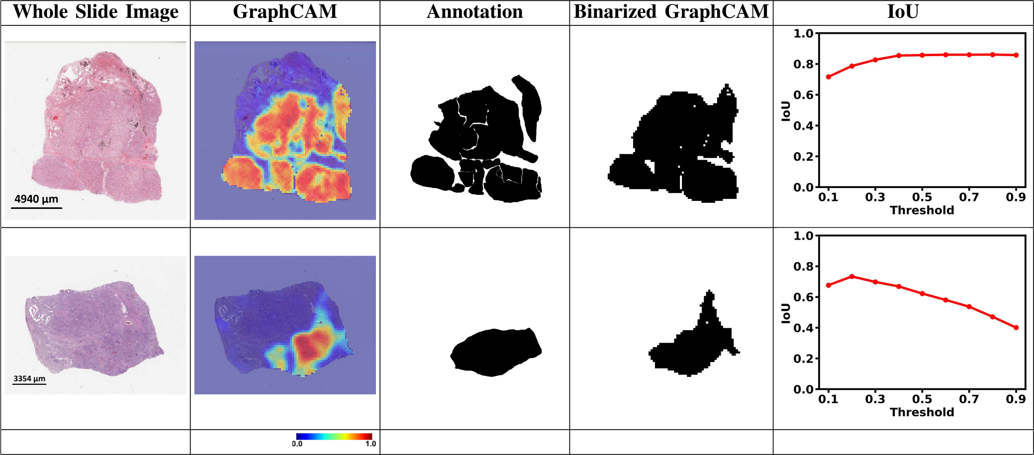
Fig. 4. GraphCAMs and their comparison with the expert annotations.

For each WSI, we generated GraphCAMs and compared them with annotations from the pathologist. The first column contains the original WSIs, the second and third columns contain GraphCAMs and pathologist’s annotations, respectively and the fourth column contains the binarized GraphCAMs based on the threshold from the Intersection of Union (IoU) plot in the last column. The first row shows an LUAD case and the second row denotes an LSCC case.
