FIGURE 2.

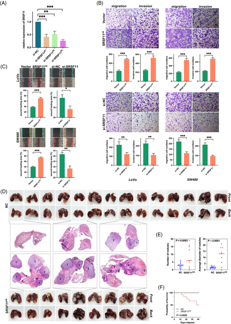
SRSF11 promotes CRC migration and invasion in vitro and metastasis in vivo. (A) qPCR analysis of SRSF11 mRNA in negative control (si‐NC) and three sets of small interfering RNA for SRSF11 (si‐SRSF11#1‐3) (Kruskal–Wallis test, **p < .01, ***p < .001). (B) Transwell assay for investigating migration and invasion capacities in SRSF11 overexpression (SRSF11OE), si‐SRSF11 as well as the negative control (Vector, si‐NC), respectively (N = 3; Student t′ test; **p < .01, ***p < .001). (C) Representative images of the cell wound healing after transfection with SRSF11OE or si‐SRSF11, as well as the corresponding negative control (N = 3; Student t′ test; *p < .05, **p < .01, ***p < .001). (D) Images of the lungs of mice 6 weeks following injections of NC and SRSF11OE group cells. The H&E staining in pulmonary metastatic foci is shown in the middle, with arrows indicating the metastatic foci. (E) Quantification of the numbers of nodules or their average diameter in both groups (N = 14 in NC group, N = 9 in SRSF11OE group; Student t test). (F) Kaplan–Meier plots of mice in NC and SRSF11OE groups (N = 15, for each, log‐rank)
