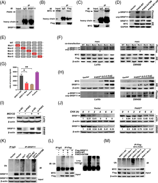
FIGURE 6.

PAK5 phosphorylates SRSF11 at serine 287 site to protect it from ubiquitination degradation. (A) Immunoblotting analysis of endogenous PAK5 protein in LoVo cells using anti‐SRSF11. (B) Immunoblotting analysis of MYC‐tag PAK5 protein in LoVo cells co‐transfected with MYC‐PAK5 and Flag‐SRSF11 plasmids using anti‐Flag. (C) Immunoblotting analysis of Flag‐tag SRSF11 protein in LoVo cells co‐transfected with MYC‐PAK5 and Flag‐SRSF11 plasmids using anti‐MYC. (D) Phos‐tag SDS‐PAGE was performed in LoVo cells after transfection with Vector, PAK5‐WT, PAK5‐K478M or PAK5‐WT+AP as illustrated in Materials and Methods. p‐SRSF11 and unp‐SRSF11 represent the phosphorylated and unphosphorylated Slug, respectively. The total protein of SRSF11 was detected by WB analysis and shown below, with GAPDH serving as an internal control. (E) Schematic diagram of SRSF11 protein, wildtype (WT) or indicated mutants (serine replaced by alanine) fused with Flag. (F) Phos‐tag SDS‐PAGE was performed in LoVo and SW480 cells after co‐transfection with MYC‐PAK5 and WT or five mutants of Flag‐SRSF11 plasmids. The asterisks represent the significant reduction of p‐SRSF11. (G) in vitro phosphorylation assay was employed to explore the effects of PAK5 on different SRSF11 and control polypeptides. WT represents the wildtype SRSF11 polypeptides with 280–294 AA sequence; Mut represents the 284 serine replaced by alanine compared with WT; random represents that the 15 amino acids are randomly arranged in disorder; positive group represents the SRSF11 polypeptides replaced by PAK5 protein (N = 5, SNK test; *p < .05, **p < .01). (H) WB analysis of SRSF11 after gradient increasing overexpression of MYC‐PAK5 or Vector plasmid in LoVo and SW480 cell lines. The Phos‐tag SDS‐PAGE was performed to detect the p‐ and unp‐SRSF11 levels as shown below. (I) The effect of PAK5 on the stability of SRSF11. MG‐132 eliminates the effect of si‐PAK5 on the stability of SRSF11. (J) LoVo and SW480 cells were treated with CHX (50 nM) for 8 h after treatment with Vector or PAK5 plasmid. SRSF11 protein levels were determined at the indicated time points (the marked number below the strip represents the relative gray value to β‐actin in the same group). (K) Immunoblotting analysis of SRSF11 protein in LoVo cells after transfection with indicated plasmids using anti‐Ub. (L) Immunoblotting analysis of Flag‐tag protein in LoVo cells after transfection with indicated plasmids using anti‐Ub. (M) Immunoblotting analysis of Flag‐tag protein in LoVo cells after transfection with indicated plasmids using anti‐Ub
