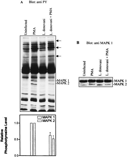
FIG. 1.
L. donovani attenuates PMA-induced tyrosine phosphorylation of MAP kinases (MAPKs). Cells were either untreated or incubated with leishmania amastigotes at an approximate parasite-to-cell ratio of 15:1. After overnight incubation (17 h), control and infected cells were incubated in the absence or presence of 100 nM PMA for 15 min. (A) Cells were lysed in modified RIPA buffer as described in Materials and Methods. Whole-cell lysates were separated by SDS-polyacrylamide gel electrophoresis (10% polyacrylamide), transferred to nitrocellulose membranes, and probed with antiphosphotyrosine (anti PY) antibodies. Blots were developed by ECL, and an autoluminogram of a blot is shown. The tyrosine-phosphorylated bands with Mr of 44,000 and 42,000 corresponded to p44MAP kinase-1 and p42MAP kinase-2, respectively. In addition to MAP kinase-1 and MAP kinase-2, the positions of other PMA-induced phosphotyrosine-containing proteins are indicated by arrows. The autoluminogram was analyzed by densitometry in the region of p44 and p42 MAP kinases. (B) The same blot was stripped and reprobed with anti-MAP kinase antibodies. The data shown are from three independent experiments that yielded similar results. The values shown in the histogram represent mean and standard deviation.

