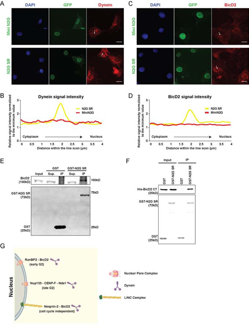
Figure 3. BicD2 interaction with Nesprin-2.
(A-D) Cultured NIH 3T3 fibroblasts were transfected with GFP-tagged Mini N2G or N2G SR cDNAs and immunostained for endogenous dynein or BicD2. Intensity scans were performed along 4μm lines through the NE (arrows). (A, C) Representative fixed images and (B, D) line scan quantifications of dynein and BicD2 signal. Cells expressing N2G SR show NE dynein and BicD2 decoration, indicating that this Nesprin fragment is sufficient to recruit both factors. (E) GST-N2G SR pull-down from embryonic rat brain lysates showing BicD2 coprecipitation. (F) Purified bacterially-expressed GST-N2G SR pulls down bacterially-expressed BicD2 CT. (G) Schematic diagram depicting multiple dynein NE recruitment mechanisms in G2 vs. Non-G2 phases of the cell cycle. During G2, dynein is recruited by two consecutive Nuclear Pore Complex-dependent pathways [13,14]. An “early pathway”, involving the nucleoporin RanBP2 and BicD2, and a “late pathway” involving the nucleoporin Nup133 and CENP-F/Nde1. In the current study we find that BicD2 and dynein are also recruited by the microtubule motor-binding domain of Nesprin-2, independent of cell cycle stage. Data are presented as superimposed symbols at mean with a connecting line in B and D. Data in B and D include line scan analysis from at least 6 and 12 cells, respectively. A and C scale bar, 10μm. Related to Figure S2.

