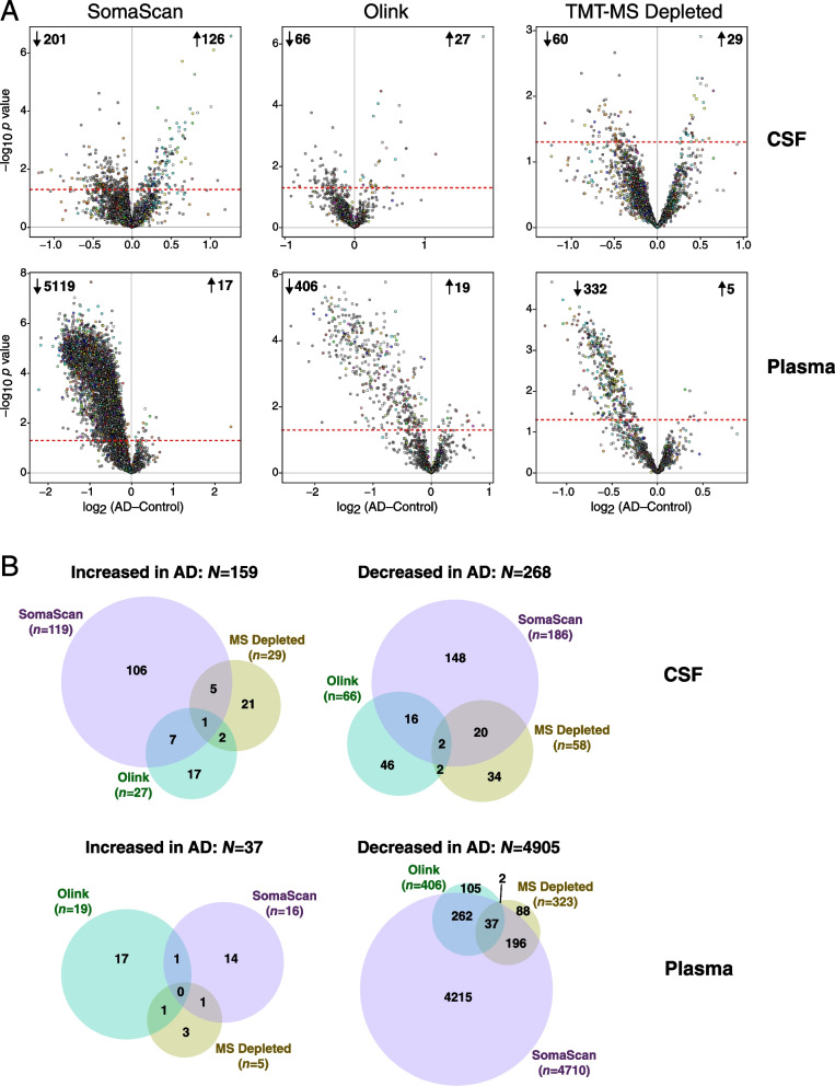
Fig. 4.
Differential protein abundance in AD by platform in CSF and plasma. A Differential protein abundance at the individual protein level between AD and control cases on the SomaScan (left), Olink (center), and TMT-MS Depleted (right) platforms, in CSF (top) and plasma (bottom). AD-control values less than 0 indicate decreased levels in AD, and values greater than 0 indicate increased levels in AD. Proteins that are above the dashed red line are significantly altered in AD by t test at p<0.05. Proteins are colored by the brain co-expression module in which they reside, as described in Johnson et al. [2] B Overlap of differential protein expression in CSF and plasma across the three platforms. Overlap includes all commonly and uniquely measured proteins in each platform and is restricted to unique gene symbols (i.e., excluding gene isoforms or redundant aptamers, which are included in A)

