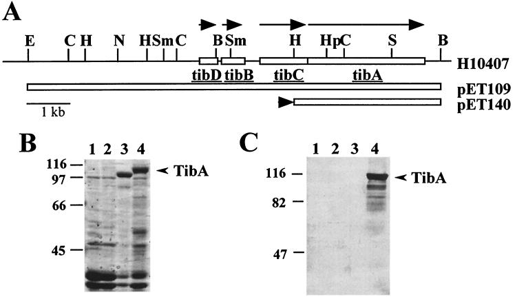
FIG. 1.
Detection of glycoproteins in outer membranes of recombinant E. coli HB101. (A) Restriction endonuclease map of the tib locus as found in the H10407 genome or in the indicated plasmids. The direction of tib gene transcription is indicated by arrows above the H10407 map. The extent of the tib locus contained by each plasmid is indicated by an open box. The black arrowhead to the left of the pET140 map indicates the direction of transcription from an exogenous promoter found in the plasmid vector. B, BamHI; C, ClaI; E, EcoRI; H, HindIII; Hp, HpaI; N, NruI; S, SalI; Sm, SmaI. (B) Coomassie blue-stained SDS-PAGE (7.5% polyacrylamide) of outer membranes purified from the following strains (by lane): 1, HB101; 2, HB101(pHC79); 3, HB101(pET140); 4, HB101(pET109). Plasmid pET140 expresses the 100-kDa preTibA protein, whereas pET109 expresses the 104-kDa TibA protein. (C) Samples identical to those shown in panel B were transferred to nitrocellulose and then stained for glycoprotein as described in Materials and Methods. The migration of molecular mass standards is shown to the left of panels B and C. The mobility of TibA is shown by an arrow to the right of panels B and C.

