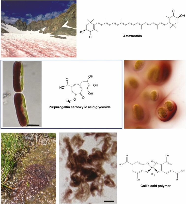
Fig. 4.
Examples of vegetative red pigmentation in terrestrial algae. The top image shows algae producing ‘blood snow’ in California, USA (USDA photograph by Paul Wade, https://commons.wikimedia.org/wiki/File:170828-FS-Inyo-PRW-001-MountRitter_(36217539154).jpg, Creative Commons Attribution 2.0 Generic licence). The middle row shows Ancylonema nordenskioeldii on the left and pigmented mucilage of a Serritaenia spp. on the right (both Zygnematophyceae). A derivative of purpurogallin has been identified as the phenolic pigment of Mesotaenium berggrenii, a relative of A. nordenskioeldii, with both these Zygnematophyceae species commonly found on alpine and Arctic glaciers and having purple-brown vacuoles containing phenolic pigments. The extracellular pigment of Serritaenia mucilage is also thought to be phenolic, but the structure has not been elucidated. The bottom row shows Zygogonium erictorum (also Zygnematophyceae), and its associated pigment thought to be a gallic acid-based polymer. Image of A. nordenskioeldii reprinted from Remias et al. (2012a). Image of Serritaenia courtesy of Dr Sebastian Hess (University of Cologne, Germany). Images of Z. erictorum reprinted from Aigner et al. (2013). Scale bars represent 10 µm (A. nordenskioeldii) and 1 cm (Z. erictorum).

