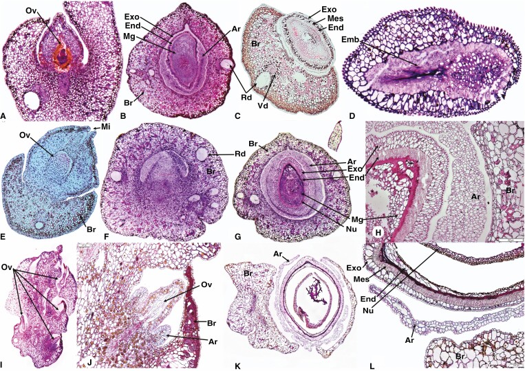
Fig. 4.
Seed cone longitudinal and cross-sections showing morpho-anatomical features of Phyllocladus trichomanoides longitudinal sections (A, B, D) and cross section (C), P. hypophyllus (E–H) and P. aspleniifolius (I–L). Br, bract; Sd= Seed ; Ov, ovule; Ar, aril; Mi, micropyle; Exo, exotesta; End, endotesta; Emb, embryo; Rd, resin duct; Vb, vascular bundles; Nu, nucellus (Nu); Mg, megagametophyte. Scale bars (H) = 200 μm; (L) = 100 μm.

