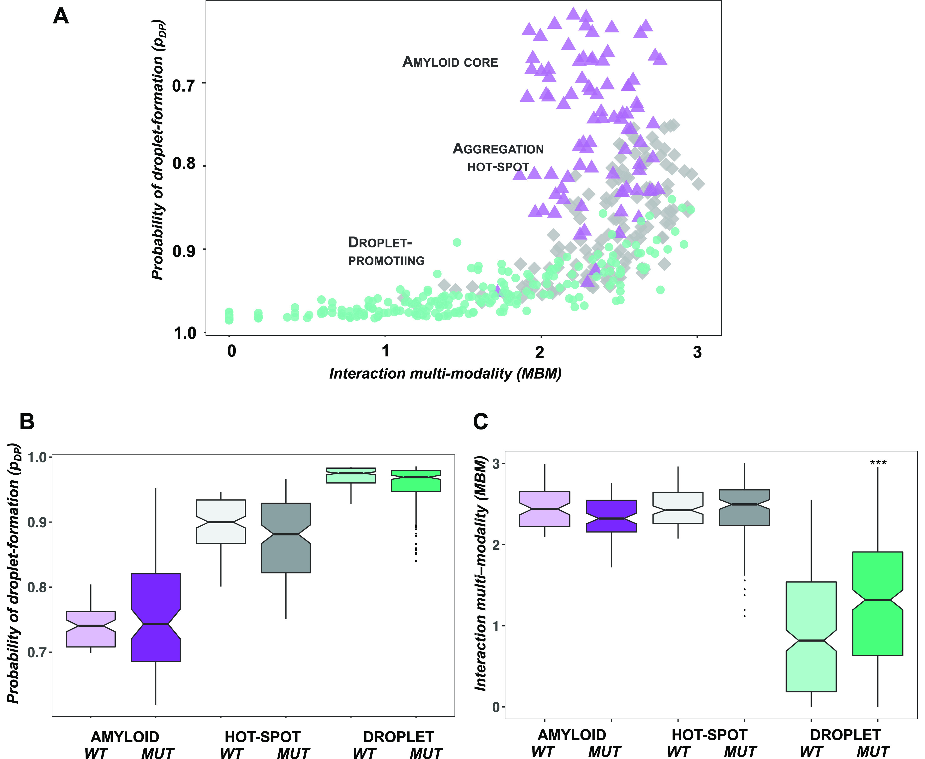
Figure 3.

Single mutations increase the MBM of the TDP-43 LC domain. We analyzed 498 single mutations with a change in cytotoxicity (Δetox) > 3σ .19 (A) Droplet landscape of TDP-43 single mutants. Amyloid core residues (residues 321–330, purple triangles) and droplet-promoting residues (residues 262–311 and 342–414, green circles) considerably overlap with the aggregation hot-spot region (residues 312–320 and 331–341, gray diamonds). This indicates a high probability to phase separate (pDP values, y axes) and a high multiplicity of binding modes (MBM, x axis) that reflects sampling both disordered and ordered interactions. (B) Comparison of droplet propensities of wild-type and mutant TDP-43 residues. No significant change was calculated between the phase separation probability of wild-type (light) and mutant residues (dark) in the amyloid core (purple), aggregation hot-spot (gray), and droplet region (green). (C) Comparison of MBM of wild-type and mutant TDP-43 residues. Mutations in the droplet region (dark green) significantly (p < 10–3) increase the MBM as compared to the wild-type values (light green), reflecting a shift in binding modes toward ordered interactions. The statistical significance was computed by the Mann–Whitney test of the R program.
