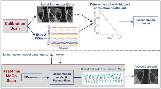
Figure 1.
Overview of the proposed prospective motion correction technique. Calibration Scan: Volunteers performed inhaling/exhaling every 5 seconds alternatively for a total of 50 seconds. The calibration data was reconstructed and processed immediately following the prescan. Kidney positions were labeled from NUFFT reconstructed images for each inhale/exhale frame. The linear motion model parameter A in Eqn. 2 was determined from the FIDnavs measured by the coil yielding the highest correlation with the kidney displacement over time. Real-time MoCo Scan: The determined linear translation parameter and coil identity number (coilmax) were used in the real-time MoCo scans. FIDnavs of coilmax was scaled and 1D Kalman-filtered in the vendor image calculation environment, and motion estimates were passed to the correction module in the sequence, which updated the applied gradients and RF pulses to compensate for translational motion.

