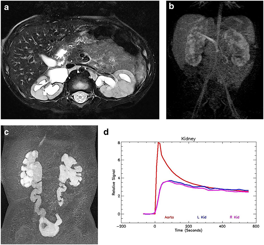
Fig. 2.
Bilateral renal dysplasia/uropathy and history of posterior urethral valves in a 1-year-old boy. a Axial T2-weighted MR image of both kidneys demonstrates mild hydronephrosis with irregular renal outlines and distortion of the renal architecture. There is no appreciable corticomedullary differentiation. b Coronal nephrographic phase after contrast administration shows bilateral irregular enhancement with no discernable concentration in the medulla. The nephrographic appearance reflects damage to the microvasculature as well as the glomeruli and tubules. c The delayed coronal post-contrast T1-weighted maximum-intensity projection (MIP) image shows bilateral hydroureteronephrosis with tortuosity of the ureters. The contrast agent eventually fills both urinary systems, although stasis and poor drainage are superimposed on the renal damage. d The signal intensity vs. time curve illustrates a normal aortic curve with bilateral dysplastic/uropathic curves for the kidneys. There is an initial brisk enhancement secondary to renal perfusion, with reduced glomerular filtration and no evidence of medullary concentration. The filtered contrast agent washed out from the renal parenchyma. The functional analysis was: vDRF L:R = 57:43; pDRF L:R = 55:45; unit Patlak L:R = 0.28:0.28 mL/min/cm3; asymmetry index = 0.0; MTT, L=44 s, R=40 s. L left, MTT mean transit time, pDRF Patlak differential renal function, R right, vDRF volumetric differential renal function.

