Fig. 3.
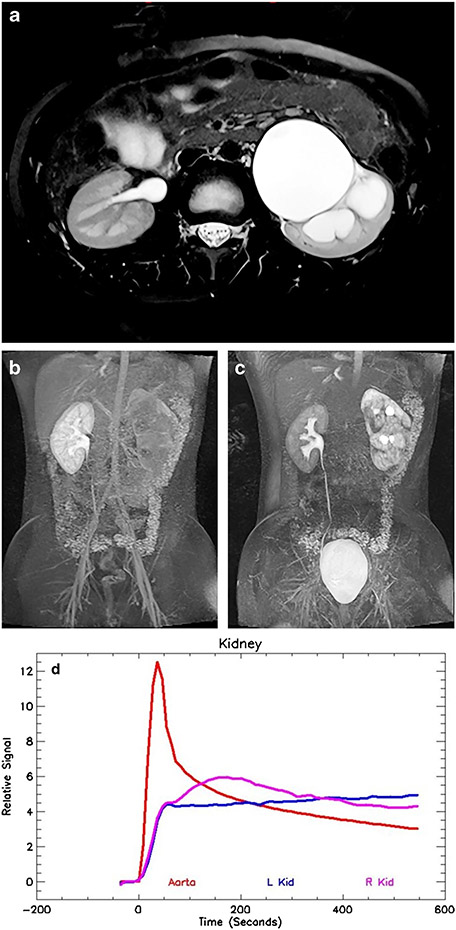
Decompensated ureteropelvic junction (UPJ) obstruction on the left in a 9-year-old boy. a Axial T2-weighted MR image shows marked dilatation and ballooning of the left renal pelvis and calyces. A small rim of medulla is still visible. Minor perinephric edema is also evident. The right kidney is normal. b Coronal late nephrographic phase maximum-intensity projection (MIP) image shows normal right kidney with prompt excretion. The left kidney has a markedly delayed and heterogeneous nephrogram. c Coronal delayed MIP shows the irregular renal outline on the left, with patchy nephrogram that is increasingly dense when compared with the normal right kidney. The increasing density is caused by a reduced glomerular filtration rate (GFR) and increased tubular reabsorption of water — the physiological responses that mitigate the effects of increased intra-pelvic pressure secondary to acute obstruction. d Signal intensity versus time curves show normal aortic and right kidney curves. The left kidney demonstrates an obstructive pattern curve with brisk enhancement secondary to renal perfusion followed by a slow and persistent increase in signal intensity reflecting parenchymal retention of the contrast medium. The renal functional analysis was: vDRF L:R = 45:55; pDRF L:R = 36:64; unit Patlak L:R = 0.12:0.21 mL/min/cm3; asymmetry index = 0.26; MTT, L=147 s, R=54 s. L left, MTT mean transit time, pDRF Patlak differential renal function, R right, vDRF volumetric differential renal function

