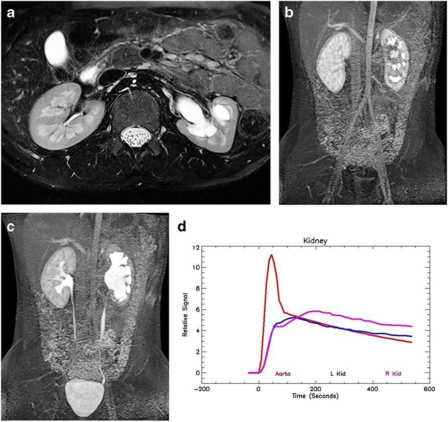
Fig. 4.
Imaging in a 9-year-old boy (same as in Fig. 3) following successful pyeloplasty. a Axial T2-weighted MR image shows significant improvement in the left-side hydronephrosis. The left kidney volume is diminished when compared to the normal right kidney, with significant loss of the renal medulla. b Coronal post-contrast maximum-intensity projection (MIP) in the nephrographic phase of the right kidney. Contrast is excreted into the calyces on the left, dramatically changed from the preoperative study. This indicates that the physiologically significant obstruction has been relieved by the pyeloplasty and that rapid calyceal excretion is largely related to a left-side concentration defect secondary to the medullary volume loss. c Coronal delayed MIP image shows contrast agent in both ureters, with a decrease in left-side hydronephrosis. Mild persistent narrowing is noted at the left ureteropelvic junction (UPJ), a not uncommon finding following pyeloplasty. d Signal intensity versus time curves again show normal aortic and right kidney curves. The left kidney curve has improved with a small concentration peak and improved washout from the renal parenchyma, with the signal paralleling the aortic curve in the later phases. The concentration peak occurred slightly earlier than in the normal right kidney, which was thought to be related to mild glomerular hyperfiltration. The renal functional analysis was: vDRF L:R = 28:72; pDRF L:R = 33:67; unit Patlak L:R = 0.33:0.26 mL/min/cm3; asymmetry index = 0.12; MTT, L=35 s, R=62 s. L left, MTT mean transit time, pDRF Patlak differential renal function, R right, vDRF volumetric differential renal function

