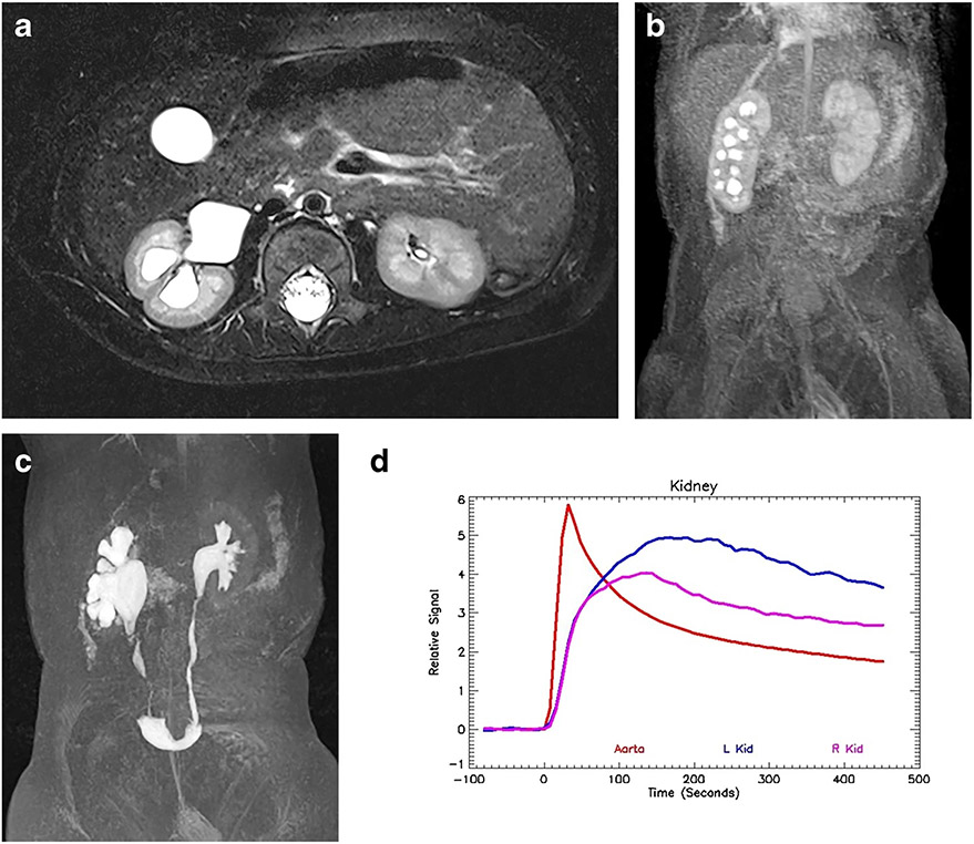
Fig. 5.
Right-side hydronephrosis but no evidence of obstruction in a 6-month-old boy. a Axial T2-weighted MR image shows moderate hydronephrosis on the right with mild loss of renal medulla. The left kidney is normal. b Coronal post-contrast maximum-intensity projection (MIP) image in the nephrographic phase of the left kidney. Contrast agent is identified in the right-side collecting system, demonstrating rapid calyceal transit time and indicating a compensated hydronephrosis. c Coronal delayed MIP image shows contrast agent filling the renal pelvis and ureters bilaterally without evidence of obstruction. d Signal intensity versus time curves show that the initial perfusion and early enhancement of the kidneys is symmetrical. However, the right kidney does not concentrate to the same degree as the left, with lower peak amplitude. There is early washout of contrast agent from the renal parenchyma, indicating an underlying tubular concentration defect and no evidence of obstruction. Correlation with the anatomical images is fundamental to interpreting the functional data. The renal functional analysis was: vDRF L:R = 56:44; pDRF L:R = 54:46; unit Patlak L:R = 0.38:0.40 mL/min/cm3; asymmetry index = 0.04; MTT, L=56 s, R=47 s. L left, MTT mean transit time, pDRF Patlak differential renal function, R right, vDRF volumetric differential renal function

