Fig. 6.
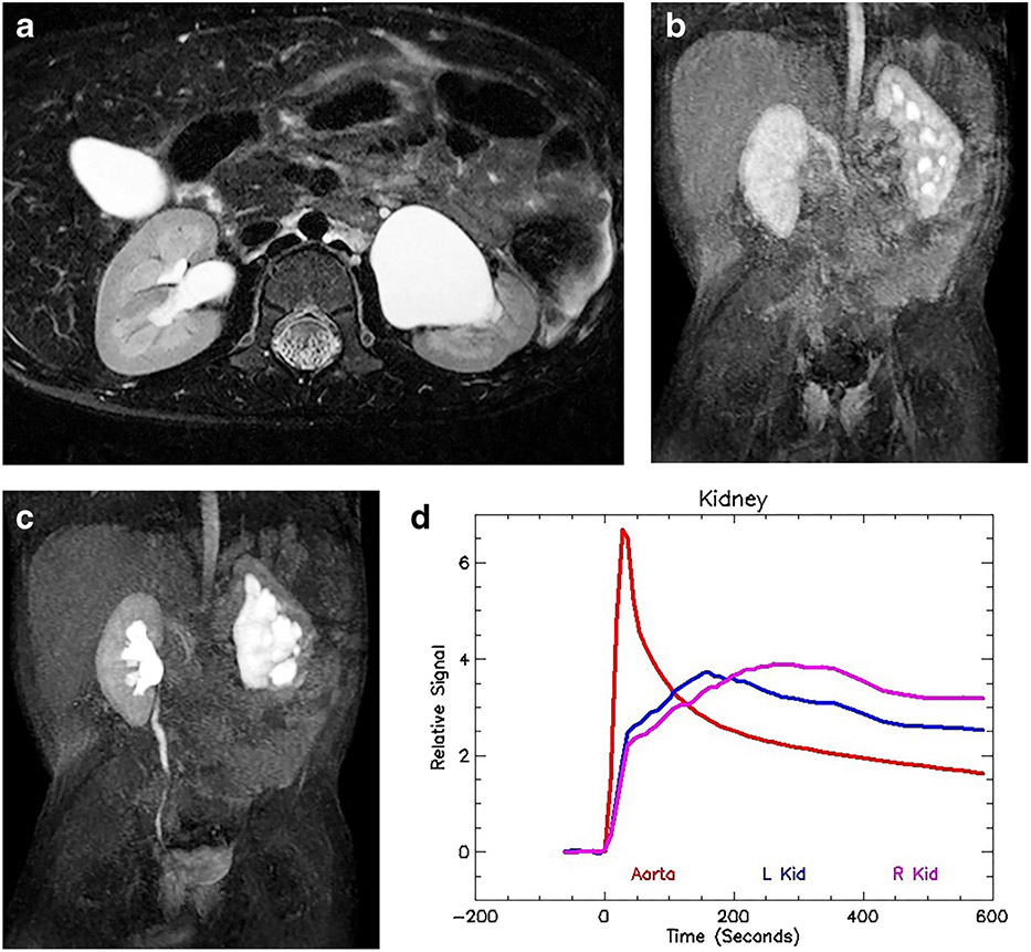
Left-side hydronephrosis and glomerular hyperfiltration in a 2-year-old boy. a Axial T2-weighted MR image shows moderate hydronephrosis on the left with mild loss of renal medulla. The right kidney is normal. b Coronal post-contrast maximum-intensity projection (MIP) image in the nephrographic phase of the right kidney. Contrast agent is identified in the left-side collecting system, demonstrating rapid calyceal transit time. c Coronal delayed MIP image shows contrast agent filling the renal pelvis bilaterally, with contrast in the right ureter. The left ureter is not seen, indicating that the renal transit time is delayed on the left as a result of stasis. The rapid calyceal excretion excludes physiologically significant obstruction. d Signal intensity versus time curves again show normal aortic and right kidney curves. The left kidney curve has a more rapid upslope with earlier peak concentration and normal washout. The whole curve for the left kidney has been shifted toward the left, indicating glomerular hyperfiltration. Glomerular hyperfiltration has been seen in cases of intermittent ureteropelvic junction (UPJ) obstruction. Without correlation with the anatomical images, it would be difficult to identify the abnormal kidney, let alone interpret the functional data. The renal functional analysis was: vDRF L:R = 47:53; pDRF L:R = 51:49; unit Patlak L:R = 0.55:0.47 mL/min/cm3; asymmetry index = 0.08; MTT, L=35 s, R=62 s. L left, MTT mean transit time, pDRF Patlak differential renal function, R right, vDRF volumetric differential renal function

