Fig. 8.
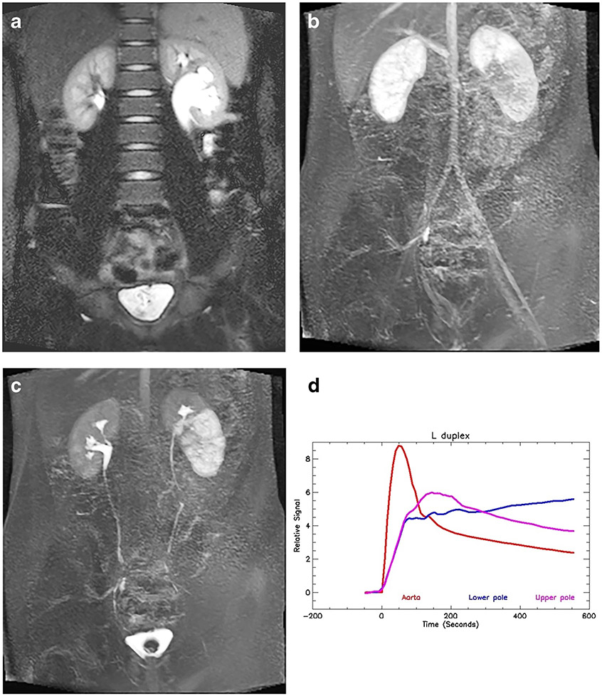
Duplex left collecting system and obstructed lower pole moiety in an 11-year-old boy. a Coronal T2-W half-Fourier acquisition single-shot turbo spin-echo (HASTE) MR image shows the duplicated left collecting system with a morphological lower-pole ureteropelvic junction (UPJ) obstruction. b Coronal post-contrast maximum-intensity projection (MIP) image from the nephrographic phase of the right kidney shows the left upper pole with a similar nephrographic appearance to (a), but the lower pole demonstrates delayed enhancement. c Coronal post-contrast T1-weighted MIP image shows normal excretion from the right kidney and the left upper pole moiety. The left lower pole moiety demonstrates a delayed and dense nephrogram with delayed calyceal transit time, indicating a physiologically significant obstruction. d Signal intensity versus time curves for the left upper- and lower-pole moieties. The upper pole curve has a normal morphology with normal parenchymal washout. The lower pole moiety shows the delayed and increasingly dense nephrogram, indicating a decompensated UPJ obstruction. The renal functional analysis was: vDRF lower:upper pole = 44:56; pDRF L:R = 36:64; unit Patlak L:R = 0.29:0.51 mL/min/cm3; asymmetry index = 0.27; MTT, upper pole = 62 s; lower pole = 142 s. L left, MTT mean transit time, pDRF Patlak differential renal function, R right, vDRF volumetric differential renal function

