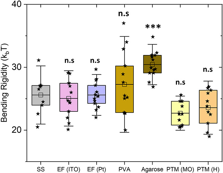
Figure 3:
Bending rigidity of bilayers measured with flickering spectroscopy of GUVs prepared by the seven GUV preparation methods. The box-plot represents the standardized distribution of data based on first quartile (Q1), mean, third quartile (Q3), and the error bars represent 1.5 Standard Deviation. The abbreviations in the figure are as follows, SS: spontaneous swelling, EF: electroformation, PVA: polyvinyl alcohol, PTM (MO): phase-transfer method (mineral oil) and PTM (H): phase-transfer method (hexadecane). The open squares represent the mean values. ANOVA comparisons test compared to spontaneous swelling which is set as control. n>10 vesicles were probed, ***p≤0.001, **p≤0.01, n.s p > 0.05.

