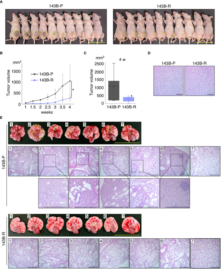
Figure 3.
Methionine-independent revertant osteosarcoma cells have reduced tumor growth and lost metastatic potential, in orthotopic xenograft mouse models. (A–C) Tumor growth in the orthotopic xenograft mouse models of 143B-P or 143B-R cells. Dashed yellow lines show the edge of the tumor tissue. Scale bar: 50 mm. *P < 0.05. (D) Representative photomicrographs of H&E-stained primary tumor tissues in the tibia of 143B-P and 143B-R cells. Scale bar: 100 μm. Magnification: 100×. (E) Spontaneous lung metastases from the tibia of 143B-P and 143B-R cells. Dashed yellow lines show metastatic lesions in the lung. Scale bar in photographs: 25 mm. Scale bar in photomicrographs: 500 μm. Magnification of photomicrographs: 40×. 143B-P: methionine-addicted parental 143B osteosarcoma cells, 143B-R: methionine-independent 143B osteosarcoma cells.

