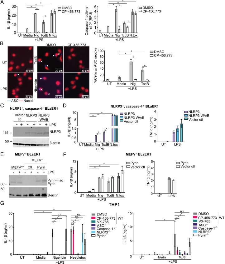
Fig 3. NLRP3, not Pyrin, is the responding inflammasome sensor to TcdB in BLaER1 cells and THP1 macrophages.
(A) Differentiated WT BLaER1 cells were primed with LPS (100 ng/ml, 3 h), preincubated with either CP-456,773 (2.5 μM, 15 min), then activated with nigericin (8 μM), TcdB (20 ng/ml), or needletox (25 ng/ml each) for 2 h. IL-1β and caspase-1 activity were assessed from the harvested supernatants. (B) ASC-mCherry transduced WT BLaER1 cells treated as in (A). ASC is in blue; nuclei are red. Cells were then fixed and the number of ASC specks quantified by microscopy. (C) NLRP3 expression in differentiated BLaER1 cells (+/− 100 ng/ml LPS, 3 h) was assessed by immunoblot. (D) Differentiated caspase-4, NLRP3 double deficient BLaER1 cells reconstituted with either NLRP3-Flag, the NLRP3 walker A/B mutant (NLRP3 WAB-Flag), or the vector control treated as in (A) and the supernatants assessed for IL-1β or TNFα. Mean and SD of 3 technical replicates shown, representative of 3 independent experiments. (E) Immunoblot of Pyrin expression in differentiated BLaER1 cells (+/− 100 ng/ml LPS, 3 h). The Pyrin-deficient cells were reconstituted with Pyrin-Flag. Representative of 3 independent experiments. (F) Differentiated Pyrin-deficient BLaER1 cells reconstituted with either Pyrin-Flag or the vector control treated as in (A) and the supernatants assessed for IL-1β or TNFα. Mean and SD of 3 technical replicates shown, representative of 3 independent experiments. (G) LPS-primed WT THP-1s or the listed KOs were activated with inflammasome activators for either 1.5 h (nigericin, needletox) or for 8 h (TcdB). Supernatants were assessed for IL-1β. Where used, CP-456,773 (2.5 μM) and VX-765 (40 μM) were preincubated with the cells for 15 min prior to addition of the inflammasome activators. Mean and SEM of 3 independent experiments shown, * p < 0.05, n.s. not significant. The underlying data can be found in the summary data file in the tab Fig 3A, 3B, 3D, 3F and 3G.

