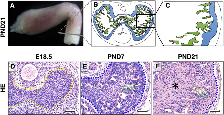
Fig. 1.
The characteristic structure of the CC during external genitalia development. (A) Lateral whole-mount image of wild-type male mouse ExG at PND21. (B, C) A schematic illustration shows the highly magnified cross section at the gray line. The region in B enclosed in the black box. (D–F) Images of HE staining at E18.5, PND7, and PND21. The yellow dotted line indicates the CC. Blue dotted lines indicate the mouse tunica albuginea. Green dotted lines indicate the mouse sinusoidal space. The asterisk indicates connective tissue. CC, corpus cavernosum; ExG, external genitalia; HE, hematoxylin and eosin; E, embryonic; PND, postnatal day. Scale bar: 500 µm.

