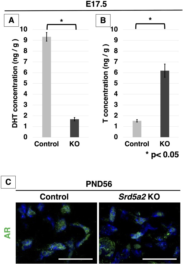
Fig. 8.
Measurement of androgens in ExG and the expression of AR in Srd5a2 KO mice. (A) Concentrations of DHT and (B) T in the CC of ExG at E17.5 measured by liquid chromatography-tandem mass spectrometry (LC-MS/MS). (C) Merged high-magnification images show the nuclear localization of AR in the control and Srd5a2 KO mouse CC at PND56. AR is present in the nucleus in both the control and Srd5a2 KO mouse sections. Scale bar in C: 100 µm. ExG, external genitalia; AR, androgen receptor; CC, corpus cavernosum; E, embryonic. Error bars represent the SEM. *P<0.05.

