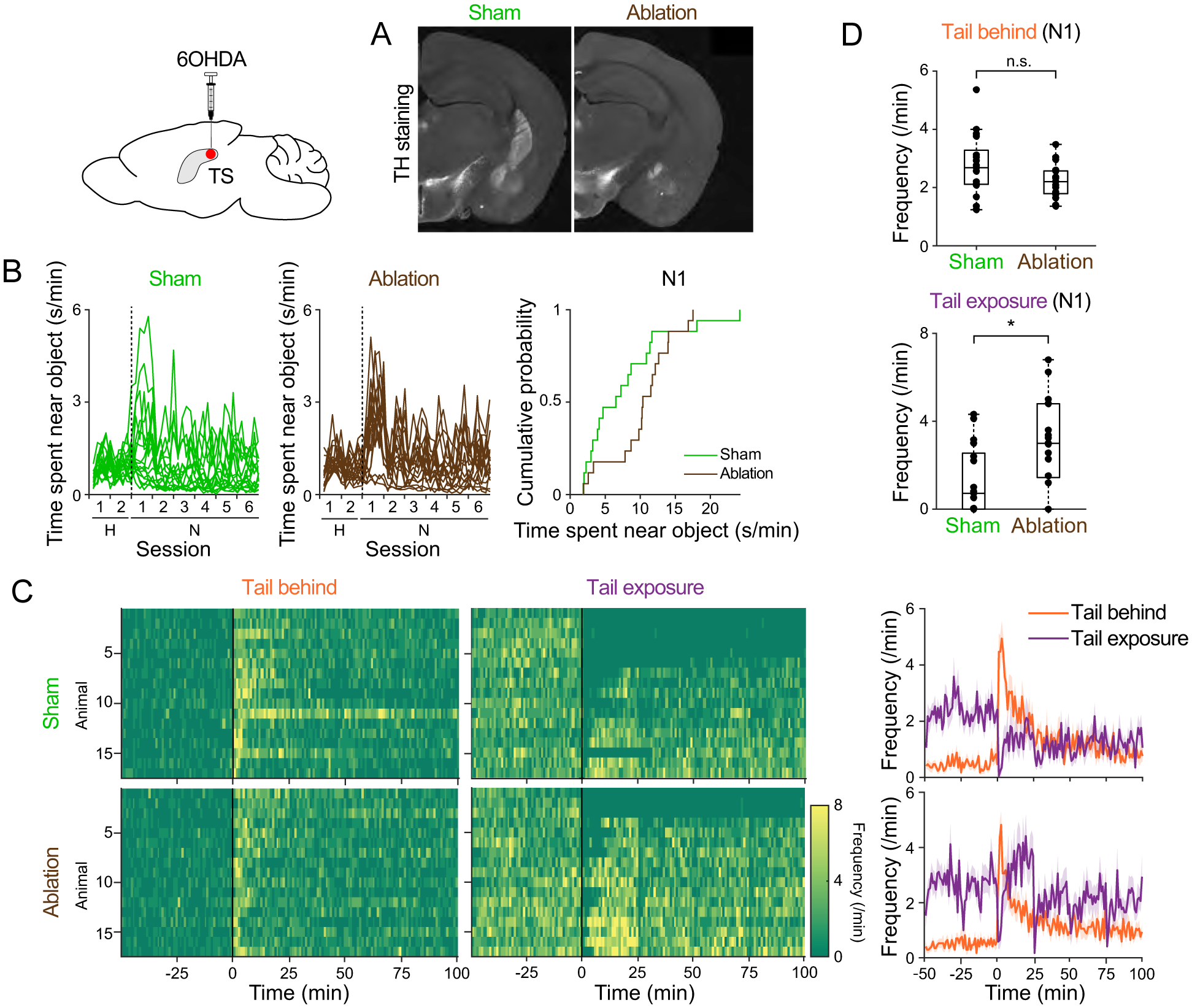
Figure 4. Ablation of TS-projecting dopamine neurons promotes post-assessment engagement.

A. Coronal sections (bregma −1.5mm) from sham (left) and ablation (right) animals. Dopamine axons were labeled with anti-tyrosine hydroxylase (TH) antibody. BLA, basolateral amygdala; CeA, central amygdala. B. Time spent near object. Right, cumulative probability on N1. Ablation vs sham, p=0.030 (K-S test). C. Frequency of each approach type bouts. Right, mean ± SEM. D. Average frequency of approach with tail behind (left; p=0.069, t-test) and approach with tail exposure (right, p=0.010, t-test) on N1. n=17 animals for each group. See also Figure S1 and Figure S2.
