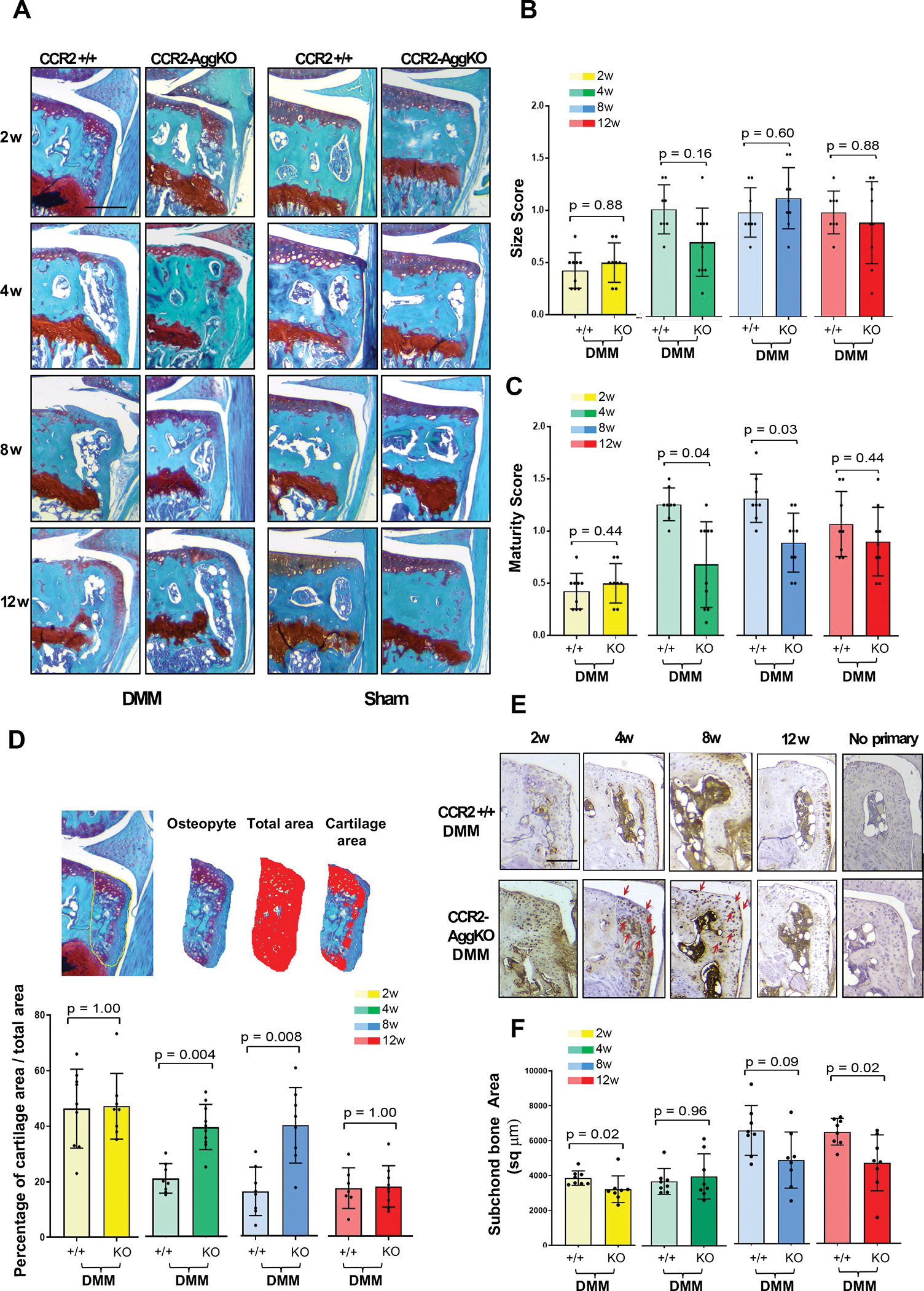
Figure 3. Osteophyte assessment in mouse knee joints of CCR2-AggKO and CCR2+/+, following early CCR2 inactivation.

(A) Safranin-O/Fast green staining of the medial compartment of CCR2-AggKO and CCR2+/+ mouse knees at 2, 4, 8 and 12 weeks after DMM surgery. Images are representative of N=8 for each of the experimental points described. (B) Osteophyte size and (C) osteophyte maturity scores (scale 0–3) of CCR2-AggKO and CCR2+/+ mouse knees at the time point indicated and described in panel A. Results are expressed as average of 4 quadrants (medial and lateral tibial plateau, medial and lateral femoral condyles); N=8 mice for each experimental point. The graphs represent the mean ± standard deviation; indicated p-values were determined by Wilcoxon rank sum tests at each time point, following adjustment for multiple comparisons. (D) Paraffin embedded knee joint sections are immunostained for Collagen 10 (Col10) at the time point indicated. Positive staining is detected as brown precipitate. Images are representative of 6 different mice for each of the experimental groups described. Scale bars of the images are 100 μm.
