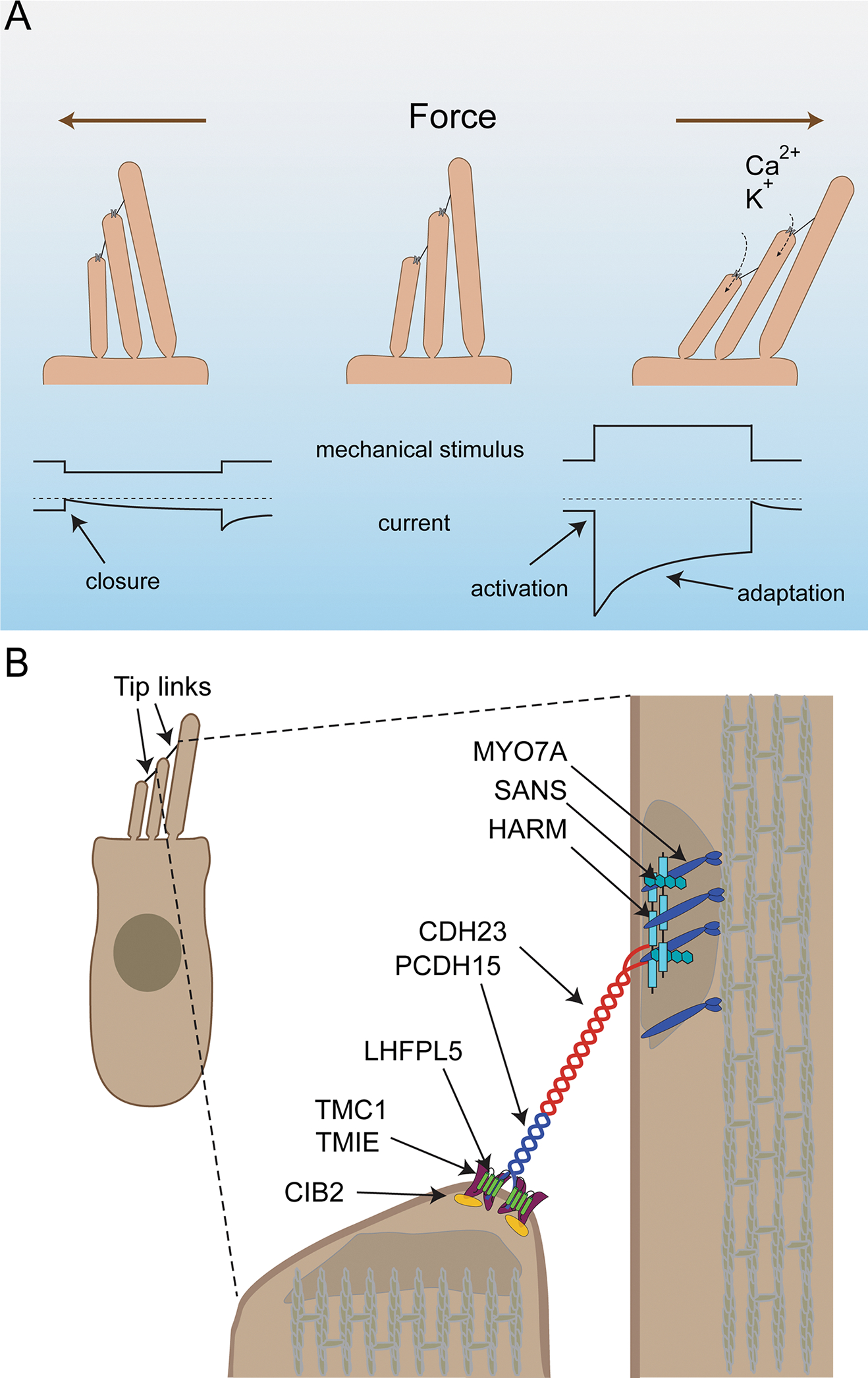
Figure 2. The mechanotransduction machinery of cochlear hair cells.

(A) Diagram of the mechanotransduction process. On top the direction of stereocilia deflection is shown, on the bottom idealized traces from recordings. Upper traces show mechanical deflections of the stereocilia: downward shift (left) indicates a deflection towards the shortest stereocilia, an upward shift (right) deflection towards the longest stereocilia. Deflection towards the shortest stereocilia (left) leads to a closing of mechanotransduction channels that are open at rest followed by slow channel re-opening. Deflection towards the longest stereocilia (right) leads to an increased influx of Ca2+ and K+ into stereocilia. This is reflected in the lower traces by a downward current, which then adapts due to channel closure even during a maintained stimulus. (B) Diagram of a hair cells indicating proteins important for mechanotransduction and their asymmetric localization at the upper and lower ends of the tip link.
